Yurdaisik Isil, Demiroz Ahu S, Oz Aysim B, Akker Mustafa, Agirman Aysegul, Aksoy Suleyman Hilmi, Nurili Fuad
Radiology, Istinye University Medical Park Gaziosmanpasa Hospital, Istanbul, TUR.
Pathology, Cerrahpaşa Faculty of Medicine, Istanbul University, Istanbul, TUR.
Cureus. 2021 Dec 27;13(12):e20734. doi: 10.7759/cureus.20734. eCollection 2021 Dec.
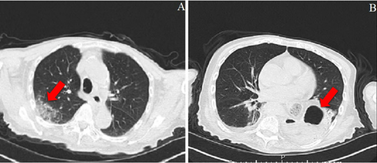
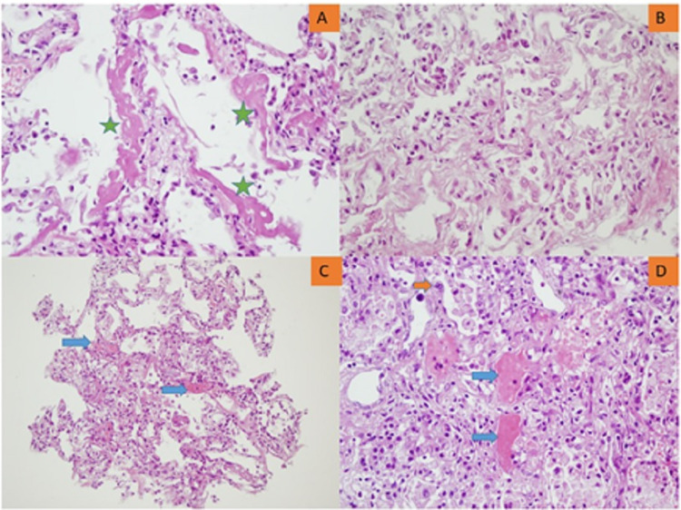
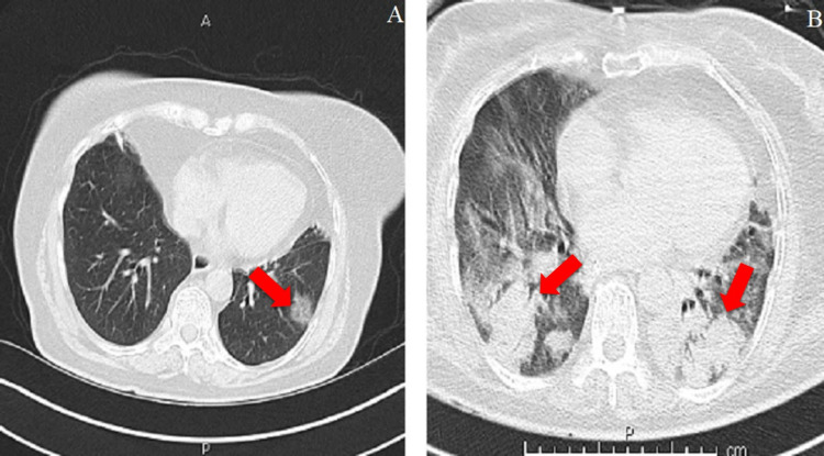
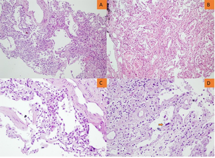
Objective We aimed to evaluate histopathologic alterations in the lung, heart, liver, and spleen of coronavirus disease 2019 (COVID-19) decedents through postmortem core needle biopsies. Materials and methods Patients who died of reverse transcription-polymerase chain reaction-proven COVID-19 were included in this postmortem case series. Postmortem percutaneous ultrasound-guided biopsies of the lungs, heart, liver, and spleen were performed using 14- and 16-gauge needles. Biopsy samples were stained with hematoxylin-eosin and examined under a light microscope. Clinicodemographic characteristics, chest computed tomography (CT) images, and COVID-19-related treatments of the patients were also collected. Results Seven patients were included in this study. Liver and heart tissue samples were available from all patients, and lung and spleen tissue samples were available from five and three patients, respectively. Chest CT images predominantly revealed bibasilar ground-glass opacities. Lung biopsies showed diffuse alveolar damage in all biopsy specimens. Heart findings were nonspecific and largely compatible with the underlying disease. Patchy necrosis, steatosis, and mononuclear cell infiltration were the main findings in the liver biopsies. Splenic histopathological examination showed that splenic necrosis and neutrophil infiltration were common findings in all patients. Conclusion Tissue acquisition was complete for the heart and liver and acceptable for the lungs. The amount of tissue was sufficient for a proper histopathologic examination. Histopathological findings were generally in accordance with previous autopsy studies. Radiological findings of the lung were also correlated with the histopathologic findings. We consider that a postmortem biopsy is a feasible alternative for histopathological examinations in COVID-19 decedents.
目的 我们旨在通过尸检粗针活检评估2019冠状病毒病(COVID-19)死者肺、心脏、肝脏和脾脏的组织病理学改变。材料与方法 本尸检病例系列纳入了经逆转录聚合酶链反应证实死于COVID-19的患者。使用14号和16号针在超声引导下对肺、心脏、肝脏和脾脏进行尸检经皮活检。活检样本用苏木精-伊红染色并在光学显微镜下检查。还收集了患者的临床人口统计学特征、胸部计算机断层扫描(CT)图像以及与COVID-19相关的治疗情况。结果 本研究纳入了7例患者。所有患者均获得了肝脏和心脏组织样本,分别有5例和3例患者获得了肺和脾脏组织样本。胸部CT图像主要显示双下肺磨玻璃影。肺活检在所有活检标本中均显示弥漫性肺泡损伤。心脏检查结果无特异性,大体上与基础疾病相符。肝脏活检的主要发现为局灶性坏死、脂肪变性和单核细胞浸润。脾脏组织病理学检查显示,脾脏坏死和中性粒细胞浸润在所有患者中均为常见表现。结论 心脏和肝脏的组织获取完整,肺部的组织获取情况可接受。组织量足以进行适当的组织病理学检查。组织病理学发现总体上与先前的尸检研究一致。肺部的影像学发现也与组织病理学发现相关。我们认为尸检活检是COVID-19死者组织病理学检查的一种可行替代方法。