• 文献检索
  • 文档翻译
  • 深度研究
  • 学术资讯
  • Suppr Zotero 插件Zotero 插件
  • 邀请有礼
  • 套餐&价格
  • 历史记录
应用&插件
Suppr Zotero 插件Zotero 插件浏览器插件Mac 客户端Windows 客户端微信小程序
定价
高级版会员购买积分包购买API积分包
服务
文献检索文档翻译深度研究API 文档MCP 服务
关于我们
关于 Suppr公司介绍联系我们用户协议隐私条款
关注我们

Suppr 超能文献

核心技术专利:CN118964589B侵权必究
粤ICP备2023148730 号-1Suppr @ 2026

文献检索

告别复杂PubMed语法,用中文像聊天一样搜索,搜遍4000万医学文献。AI智能推荐,让科研检索更轻松。

立即免费搜索

文件翻译

保留排版,准确专业,支持PDF/Word/PPT等文件格式,支持 12+语言互译。

免费翻译文档

深度研究

AI帮你快速写综述,25分钟生成高质量综述,智能提取关键信息,辅助科研写作。

立即免费体验

新型冠状病毒肺炎的尸检胸部计算机断层扫描:一种微创尸检方法

Postmortem chest computed tomography in COVID-19: A minimally invasive autopsy method.

作者信息

Savoia Paulo, Valente Yamada Sawamura Marcio, de Almeida Monteiro Renata Aparecida, Nunes Duarte-Neto Amaro, Morais Martin Maria da Graça, Dolhnikoff Marisa, Mauad Thais, Nascimento Saldiva Paulo Hilário, da Costa Leite Claudia, Ferraz da Silva Luiz Fernando, Cardoso Ellison Fernando

机构信息

Department of Radiology, University of Sao Paulo School of Medicine, Institute of Radiology, Rua Doutor Ovidio Pires de Campos, 75, 05403-010, Cerqueira Cesar, São Paulo, SP, Brazil.

Department of Pathology, University of Sao Paulo School of Medicine, Av. Dr. Arnaldo, 455, sala 1155, 01246-903, Cerqueira Cesar, São Paulo, SP, Brazil.

出版信息

Eur J Radiol Open. 2024 Jan 13;12:100546. doi: 10.1016/j.ejro.2024.100546. eCollection 2024 Jun.

DOI:10.1016/j.ejro.2024.100546
PMID:38293283
原文链接:https://pmc.ncbi.nlm.nih.gov/articles/PMC10825618/
Abstract

OBJECTIVES

Performing autopsies in a pandemic scenario is challenging, as the need to understand pathophysiology must be balanced with the contamination risk. A minimally invasive autopsy might be a solution. We present a model that combines radiology and pathology to evaluate postmortem CT lung findings and their correlation with histopathology.

METHODS

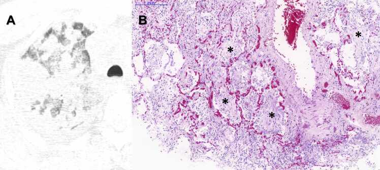
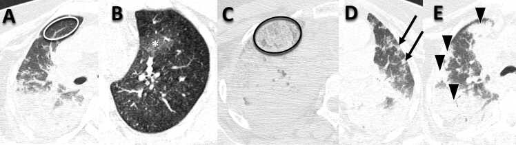
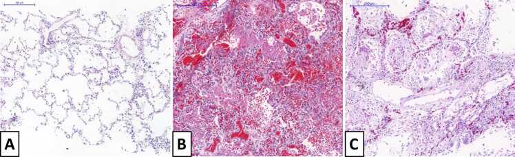
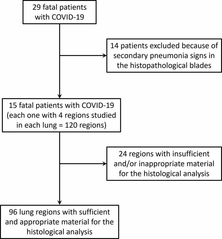
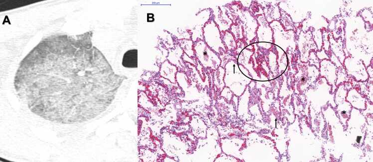
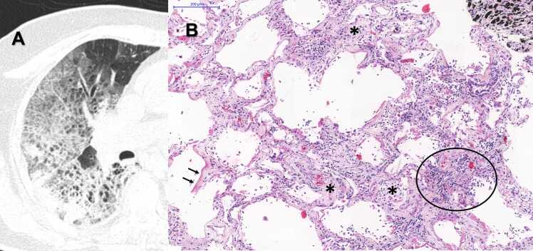
Twenty-nine patients with fatal COVID-19 underwent postmortem chest CT, and multiple lung tissue samples were collected. The chest CT scans were analyzed and quantified according to lung involvement in five categories: normal, ground-glass opacities, crazy-paving, small consolidations, and large or lobar consolidations. The lung tissue samples were examined and quantified in three categories: normal lung, exudative diffuse alveolar damage (DAD), and fibroproliferative DAD. A linear index was used to estimate the global severity of involvement by CT and histopathological analysis.

RESULTS

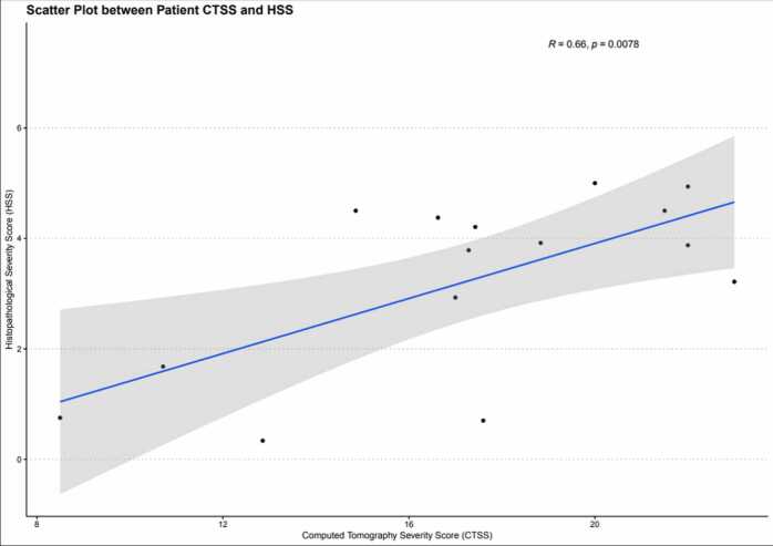
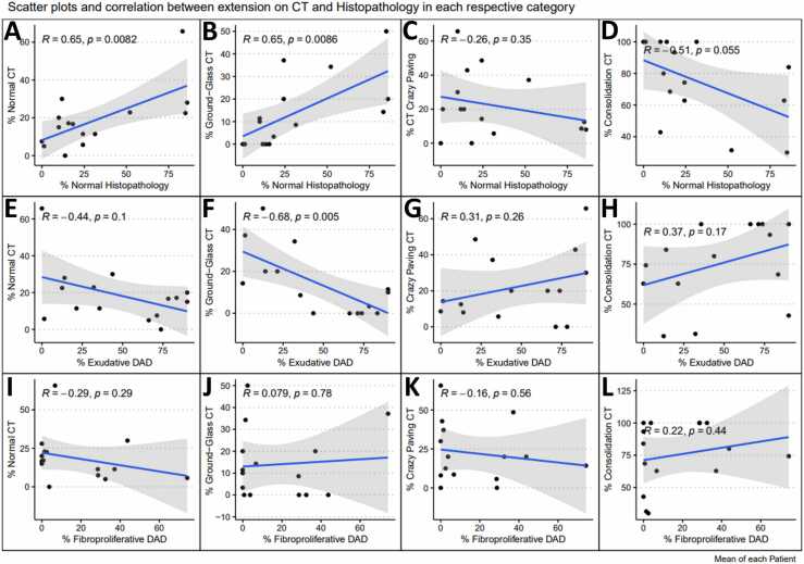
There was a positive correlation between patient mean CT and histopathological severity score indexes - Pearson correlation coefficient (R) = 0.66 (p = 0.0078). When analyzing the mean lung involvement percentage of each finding, positive correlations were found between the normal lung percentage between postmortem CT and histopathology (R=0.65, p = 0.0082), as well as between ground-glass opacities in postmortem CT and normal lungs in histopathology (R=0.65, p = 0.0086), but negative correlations were observed between ground-glass opacities extension and exudative diffuse alveolar damage in histological slides (R=-0.68, p = 0.005). Additionally, it was found is a trend toward a decrease in the percentage of normal lung tissue on the histological slides as the percentage of consolidations in postmortem CT scans increased (R =-0.51, p = 0.055). The analysis of the other correlations between the percentage of each finding did not show any significant correlation or correlation trends (p ≥ 0.10).

CONCLUSIONS

A minimally invasive autopsy is valid. As the severity of involvement is increased in CT, more advanced disease is seen on histopathology. However, we cannot state that one specific radiological category represents a specific pathological correspondent. Ground-glass opacities, in the postmortem stage, must be interpreted with caution, as expiratory lungs may overestimate disease.

摘要

目的

在大流行情况下进行尸检具有挑战性,因为理解病理生理学的需求必须与污染风险相平衡。微创尸检可能是一种解决方案。我们提出了一种结合放射学和病理学的模型,以评估死后胸部CT肺部表现及其与组织病理学的相关性。

方法

对29例死于COVID-19的患者进行了死后胸部CT检查,并采集了多个肺组织样本。根据肺部受累情况将胸部CT扫描分为五类进行分析和量化:正常、磨玻璃影、铺路石征、小实变和大或大叶实变。对肺组织样本进行检查并分为三类进行量化:正常肺、渗出性弥漫性肺泡损伤(DAD)和纤维增生性DAD。使用线性指数通过CT和组织病理学分析估计受累的总体严重程度。

结果

患者平均CT严重程度评分指数与组织病理学严重程度评分指数之间存在正相关——Pearson相关系数(R)=0.66(p=0.0078)。分析每种表现的平均肺受累百分比时,发现死后CT与组织病理学中正常肺百分比之间存在正相关(R=0.65,p=0.0082),死后CT中的磨玻璃影与组织病理学中的正常肺之间也存在正相关(R=0.65,p=0.0086),但组织学切片中磨玻璃影范围与渗出性弥漫性肺泡损伤之间存在负相关(R=-0.68,p=0.005)。此外,发现随着死后CT扫描实变百分比的增加,组织学切片中正常肺组织百分比有下降趋势(R=-0.51,p=0.055)。对每种表现百分比之间的其他相关性分析未显示任何显著相关性或相关趋势(p≥0.10)。

结论

微创尸检是有效的。随着CT中受累严重程度增加,组织病理学上可见更严重的疾病。然而,我们不能说某一特定的放射学类别代表特定的病理学对应类型。在死后阶段,磨玻璃影必须谨慎解读,因为呼气时的肺部情况可能高估疾病。

https://cdn.ncbi.nlm.nih.gov/pmc/blobs/d967/10825618/77196b0b0c5a/gr10.jpg
https://cdn.ncbi.nlm.nih.gov/pmc/blobs/d967/10825618/6afa2c6043d4/gr1.jpg
https://cdn.ncbi.nlm.nih.gov/pmc/blobs/d967/10825618/3af786ea56f0/gr2.jpg
https://cdn.ncbi.nlm.nih.gov/pmc/blobs/d967/10825618/4f895434daba/gr3.jpg
https://cdn.ncbi.nlm.nih.gov/pmc/blobs/d967/10825618/e4c5b02e8d5e/gr4.jpg
https://cdn.ncbi.nlm.nih.gov/pmc/blobs/d967/10825618/444af0653df5/gr5.jpg
https://cdn.ncbi.nlm.nih.gov/pmc/blobs/d967/10825618/ce6faeb77f89/gr6.jpg
https://cdn.ncbi.nlm.nih.gov/pmc/blobs/d967/10825618/8d0fbc92484d/gr7.jpg
https://cdn.ncbi.nlm.nih.gov/pmc/blobs/d967/10825618/f1ac5bc14ab7/gr8.jpg
https://cdn.ncbi.nlm.nih.gov/pmc/blobs/d967/10825618/ed5bc184b918/gr9.jpg
https://cdn.ncbi.nlm.nih.gov/pmc/blobs/d967/10825618/77196b0b0c5a/gr10.jpg

相似文献

1
Postmortem chest computed tomography in COVID-19: A minimally invasive autopsy method.新型冠状病毒肺炎的尸检胸部计算机断层扫描:一种微创尸检方法
Eur J Radiol Open. 2024 Jan 13;12:100546. doi: 10.1016/j.ejro.2024.100546. eCollection 2024 Jun.
2
Postmortem CT pulmonary findings in SARS-CoV-2-positive cases: correlation with lung histopathological findings and autopsy results.COVID-19 逝者的 CT 肺部影像表现:与肺组织病理发现及尸检结果的相关性。
Int J Legal Med. 2022 Sep;136(5):1407-1415. doi: 10.1007/s00414-022-02793-2. Epub 2022 Feb 14.
3
COVID-19 Infection in Octagenarian Patients: Imaging Aspects and Clinical Correlations.八旬老人感染 COVID-19:影像学表现及临床相关性。
Gerontology. 2022;68(3):261-271. doi: 10.1159/000516166. Epub 2021 Aug 5.
4
Radio-Histological Correlation of Lung Features in Severe COVID-19 Through CT-Scan and Lung Ultrasound Evaluation.通过CT扫描和肺部超声评估对重症COVID-19患者肺部特征进行放射学与组织学相关性研究
Front Med (Lausanne). 2022 Apr 19;9:820661. doi: 10.3389/fmed.2022.820661. eCollection 2022.
5
Correlation of autopsy pathological findings and imaging features from 9 fatal cases of COVID-19 pneumonia.COVID-19 肺炎 9 例死亡病例尸检病理发现与影像学特征的相关性。
Medicine (Baltimore). 2021 Mar 26;100(12):e25232. doi: 10.1097/MD.0000000000025232.
6
Radiological Patterns of Lung Injury on HRCT in COVID-19 Patients: An Experience From Tertiary Care Centre in India.新型冠状病毒肺炎患者高分辨率CT上肺损伤的影像学表现:来自印度一家三级医疗中心的经验
Cureus. 2023 Nov 7;15(11):e48479. doi: 10.7759/cureus.48479. eCollection 2023 Nov.
7
Time Course of Lung Changes at Chest CT during Recovery from Coronavirus Disease 2019 (COVID-19).新冠肺炎(COVID-19)康复过程中胸部 CT 肺部变化的时间进程。
Radiology. 2020 Jun;295(3):715-721. doi: 10.1148/radiol.2020200370. Epub 2020 Feb 13.
8
COVID-19: Histopathological correlates of imaging patterns on chest computed tomography.COVID-19:胸部计算机断层扫描成像模式的组织病理学相关性。
Respirology. 2021 Sep;26(9):869-877. doi: 10.1111/resp.14101. Epub 2021 Jun 22.
9
Late histopathologic characteristics of critically ill COVID-19 patients: Different phenotypes without evidence of invasive aspergillosis, a case series.危重症 COVID-19 患者的晚期组织病理学特征:无侵袭性曲霉菌病证据的不同表型,一项病例系列研究。
J Crit Care. 2020 Oct;59:149-155. doi: 10.1016/j.jcrc.2020.07.002. Epub 2020 Jul 8.
10
Ultrasound assessment of pulmonary fibroproliferative changes in severe COVID-19: a quantitative correlation study with histopathological findings.超声评估重症 COVID-19 肺纤维化增生性改变:与组织病理学发现的定量相关性研究。
Intensive Care Med. 2021 Feb;47(2):199-207. doi: 10.1007/s00134-020-06328-4. Epub 2021 Jan 3.

本文引用的文献

1
Performance of an AI algorithm during the different phases of the COVID pandemics: what can we learn from the AI and vice versa.人工智能算法在新冠疫情不同阶段的表现:我们能从人工智能中学到什么,反之亦然。
Eur J Radiol Open. 2023 Dec;11:100497. doi: 10.1016/j.ejro.2023.100497. Epub 2023 Jun 19.
2
Postmortem CT pulmonary findings in SARS-CoV-2-positive cases: correlation with lung histopathological findings and autopsy results.COVID-19 逝者的 CT 肺部影像表现:与肺组织病理发现及尸检结果的相关性。
Int J Legal Med. 2022 Sep;136(5):1407-1415. doi: 10.1007/s00414-022-02793-2. Epub 2022 Feb 14.
3
Postmortem Biopsies of the Lung, Heart, Liver, and Spleen of COVID-19 Patients.
新冠病毒肺炎患者肺、心、肝、脾的尸检活检
Cureus. 2021 Dec 27;13(12):e20734. doi: 10.7759/cureus.20734. eCollection 2021 Dec.
4
Postmortem Chest Computed Tomography in Fatal COVID-19: A Valuable Diagnostic Tool for Minimally Invasive Autopsy.致命性新冠肺炎的尸检胸部计算机断层扫描:一种用于微创尸检的有价值诊断工具。
Clinics (Sao Paulo). 2021 Dec 8;76:e3551. doi: 10.6061/clinics/2021/e3551. eCollection 2021.
5
A case of persistent severe sequelae of COVID-19 infection: potential role in sudden death?新冠肺炎感染持续严重后遗症 1 例:是否与猝死有关?
Forensic Sci Med Pathol. 2022 Mar;18(1):69-73. doi: 10.1007/s12024-021-00435-4. Epub 2021 Oct 19.
6
Dying "from" or "with" COVID-19 during the Pandemic: Medico-Legal Issues According to a Population Perspective.大流行期间死于或伴有 COVID-19:从人口角度看医学法律问题。
Int J Environ Res Public Health. 2021 Aug 22;18(16):8851. doi: 10.3390/ijerph18168851.
7
Post-mortem CT lung findings at a medicolegal institute in SARS-CoV-2 RT-PCR positive cases with autopsy correlation.SARS-CoV-2 RT-PCR 阳性病例尸检相关性分析:法医学研究所中死后 CT 肺部检查结果。
Forensic Sci Med Pathol. 2021 Dec;17(4):611-620. doi: 10.1007/s12024-021-00389-7. Epub 2021 Jun 23.
8
Use of post-mortem chest computed tomography in Covid-19 pneumonia.死后胸部计算机断层扫描在新冠肺炎肺炎中的应用。
Forensic Sci Int. 2021 Aug;325:110851. doi: 10.1016/j.forsciint.2021.110851. Epub 2021 May 27.
9
Role of High Resolution Computed Tomography chest in the diagnosis and evaluation of COVID -19 patients -A systematic review and meta-analysis.胸部高分辨率计算机断层扫描在2019冠状病毒病患者诊断和评估中的作用——一项系统评价和荟萃分析。
Eur J Radiol Open. 2021;8:100350. doi: 10.1016/j.ejro.2021.100350. Epub 2021 May 13.
10
Postmortem radiologic and pathologic findings in COVID-19: The Toronto experience with pre-hospitalization deaths in the community.COVID-19 死后放射学和病理学表现:多伦多社区中院前死亡的经验。
Forensic Sci Int. 2021 May;322:110755. doi: 10.1016/j.forsciint.2021.110755. Epub 2021 Mar 17.