Institute of Veterinary Anatomy, Histology and Embryology, Justus-Liebig-University of Giessen, Frankfurter Str. 98, 35392, Giessen, Germany.
Anatomy and Embryology Department, Faculty of Veterinary Medicine, University of Mansoura, Mansoura, 35516, Egypt.
Stem Cell Res Ther. 2022 Feb 5;13(1):56. doi: 10.1186/s13287-022-02730-5.
Skeletal muscle-derived stem cells (SC) have become a promising approach for investigating myogenic differentiation and optimizing tissue regeneration. Muscle regeneration is performed by SC, a self-renewal cell population underlying the basal lamina of muscle fibers. Here, we examined the impact of hypoxia condition on the regenerative capacity of SC either in their native microenvironment or via isolation in a monolayer culture using ectopic differentiation inductions. Furthermore, the effect of low oxygen tension on myogenic differentiation protocols of the myoblasts cell line C2C12 was examined.
Hind limb muscles of wild type mice were processed for both SC/fiber isolation and myoblast extraction using magnetic beads. SC were induced for myogenic, adipogenic and osteogenic commitments under normoxic (21% O) and hypoxic (3% O) conditions. SC proliferation and differentiation were evaluated using histological staining, immunohistochemistry, morphometric analysis and RT-qPCR. The data were statistically analyzed using ANOVA.
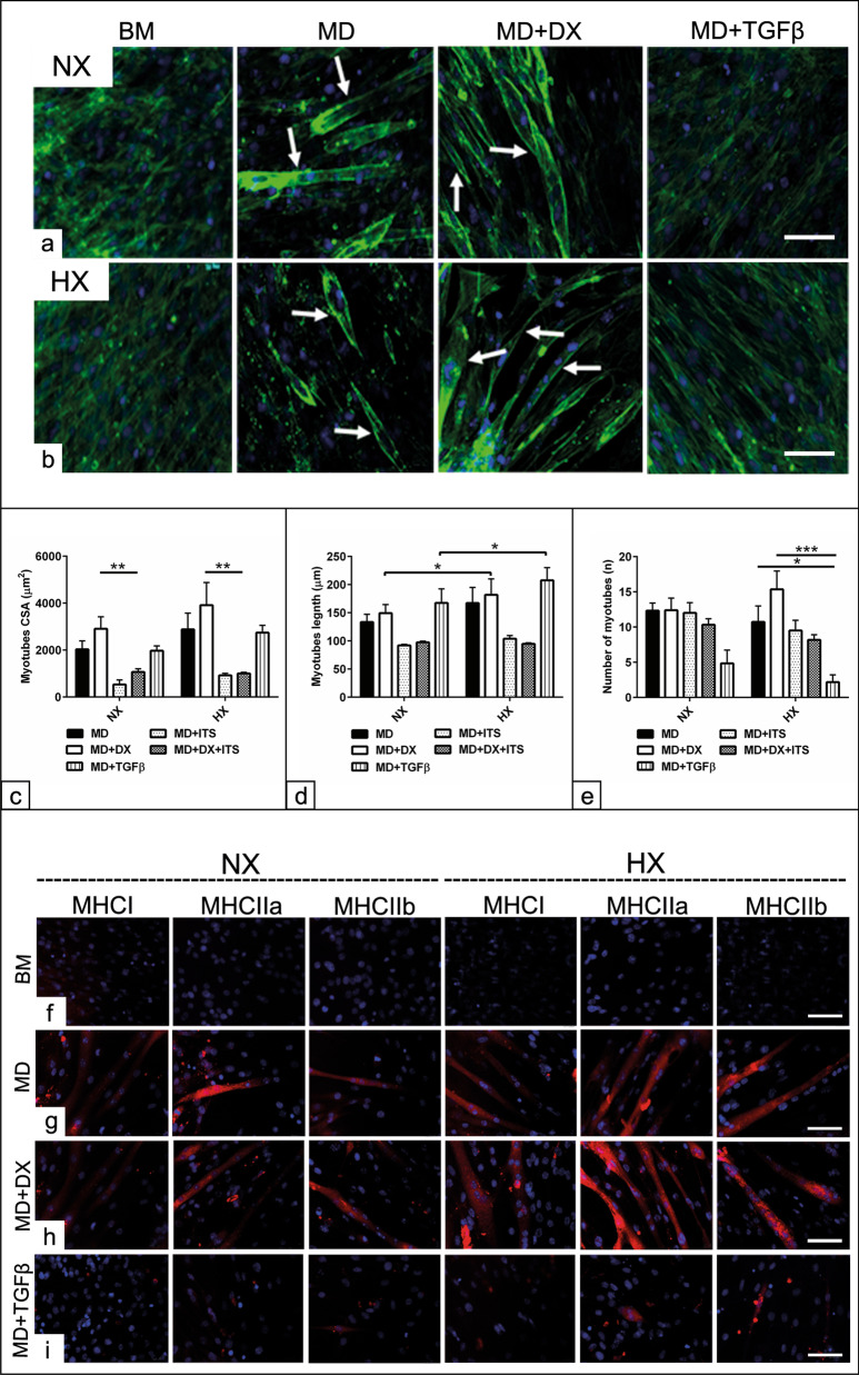
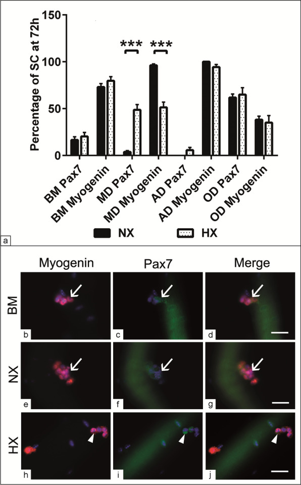
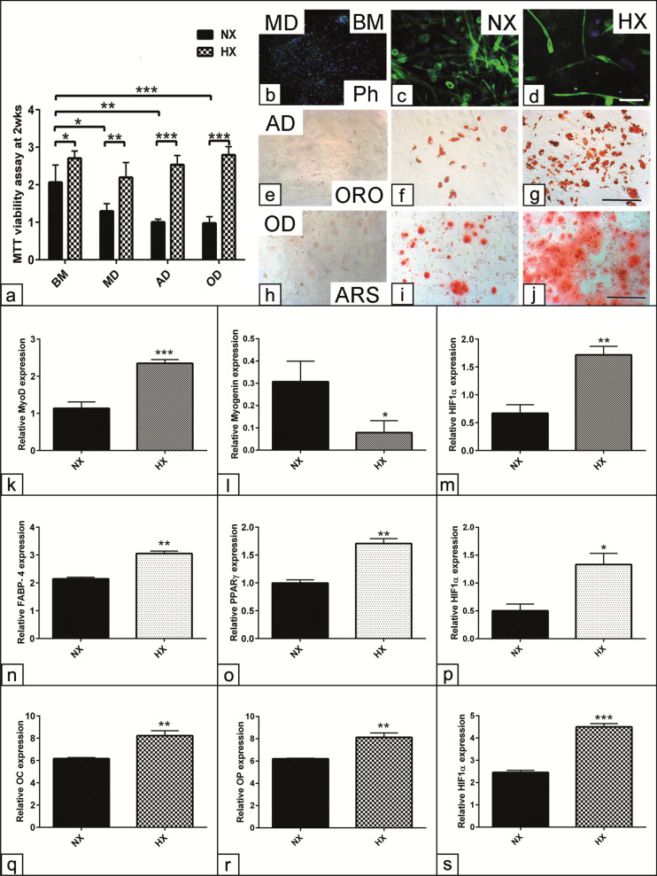
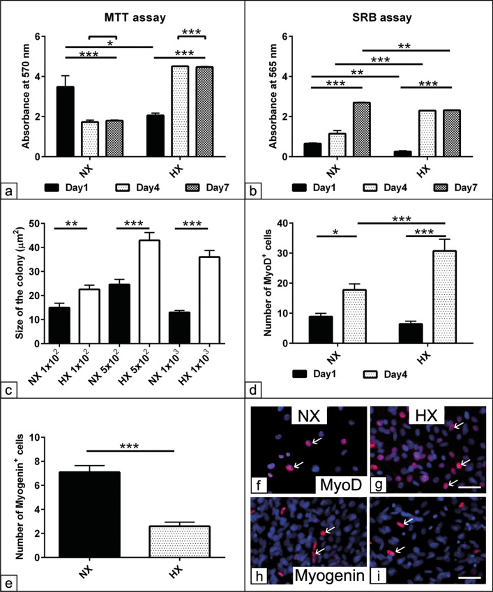
The data revealed enhanced SC proliferation and motility following differentiation induction after 48 h under hypoxia. Following myogenic induction, the number of undifferentiated cells positive for Pax7 were increased at 72 h under hypoxia. Hypoxia upregulated MyoD and downregulated Myogenin expression at day-7 post-myogenic induction. Hypoxia promoted both SC adipogenesis and osteogenesis under respective induction as shown by using Oil Red O and Alizarin Red S staining. The expression of adipogenic markers; peroxisome proliferator activated receptor gamma (PPARγ) and fatty acid-binding protein 4 (FABP4) were upregulated under hypoxia up to day 14 compared to normoxic condition. Enhanced osteogenic differentiation was detected under hypoxic condition via upregulation of osteocalcin and osteopontin expression up to day 14 as well as, increased calcium deposition at day 21. Hypoxia exposure increases the number of adipocytes and the size of fat vacuoles per adipocyte compared to normoxic culture. Combining the differentiation medium with dexamethasone under hypoxia improves the efficiency of the myogenic differentiation protocol of C2C12 by increasing the length of the myotubes.
Hypoxia exposure increases cell resources for clinical applications and promotes SC multipotency and thus beneficial for tissue regeneration.
骨骼肌源性干细胞 (SC) 已成为研究成肌分化和优化组织再生的有前途的方法。SC 是一种自我更新的细胞群体,位于肌肉纤维的基底层,它可以进行肌肉再生。在这里,我们研究了缺氧条件对 SC 在其天然微环境中或通过在单层培养中进行异位分化诱导进行分离时的再生能力的影响。此外,还研究了低氧张力对成肌细胞系 C2C12 的成肌分化方案的影响。
使用磁珠从野生型小鼠的后肢肌肉中处理 SC/纤维分离和成肌细胞提取。在常氧 (21% O) 和缺氧 (3% O) 条件下诱导 SC 进行成肌、成脂和成骨分化。使用组织学染色、免疫组织化学、形态计量分析和 RT-qPCR 评估 SC 的增殖和分化。使用方差分析对数据进行统计学分析。
数据显示,缺氧 48 小时后诱导分化时,SC 的增殖和迁移能力增强。在成肌诱导后,72 小时时缺氧下 Pax7 阳性的未分化细胞数量增加。缺氧诱导后第 7 天,MyoD 上调,Myogenin 表达下调。缺氧条件下促进 SC 成脂和成骨分化,用油红 O 和茜素红 S 染色显示。在缺氧条件下,脂肪细胞分化标志物过氧化物酶体增殖物激活受体 γ (PPARγ) 和脂肪酸结合蛋白 4 (FABP4) 的表达在第 14 天之前上调,与常氧条件相比。在第 14 天之前,通过骨钙素和骨桥蛋白表达的上调以及第 21 天钙沉积的增加,在缺氧条件下检测到增强的成骨分化。与常氧培养相比,缺氧暴露会增加脂肪细胞的数量和每个脂肪细胞的脂肪空泡大小。在缺氧条件下将分化培养基与地塞米松结合使用可通过增加肌管的长度来提高 C2C12 的成肌分化方案的效率。
缺氧暴露增加了用于临床应用的细胞资源,并促进了 SC 的多能性,从而有益于组织再生。