• 文献检索
  • 文档翻译
  • 深度研究
  • 学术资讯
  • Suppr Zotero 插件Zotero 插件
  • 邀请有礼
  • 套餐&价格
  • 历史记录
应用&插件
Suppr Zotero 插件Zotero 插件浏览器插件Mac 客户端Windows 客户端微信小程序
定价
高级版会员购买积分包购买API积分包
服务
文献检索文档翻译深度研究API 文档MCP 服务
关于我们
关于 Suppr公司介绍联系我们用户协议隐私条款
关注我们

Suppr 超能文献

核心技术专利:CN118964589B侵权必究
粤ICP备2023148730 号-1Suppr @ 2026

文献检索

告别复杂PubMed语法,用中文像聊天一样搜索,搜遍4000万医学文献。AI智能推荐,让科研检索更轻松。

立即免费搜索

文件翻译

保留排版,准确专业,支持PDF/Word/PPT等文件格式,支持 12+语言互译。

免费翻译文档

深度研究

AI帮你快速写综述,25分钟生成高质量综述,智能提取关键信息,辅助科研写作。

立即免费体验

巩膜压迫辅助下的周边光学相干断层扫描在早产儿视网膜病变中的应用

Peripheral OCT Assisted by Scleral Depression in Retinopathy of Prematurity.

作者信息

Scruggs Brittni A, Ni Shuibin, Nguyen Thanh-Tin P, Ostmo Susan, Chiang Michael F, Jia Yali, Huang David, Jian Yifan, Campbell J Peter

机构信息

Casey Eye Institute, Department of Ophthalmology, Oregon Health & Science University, Portland, OR, USA.

National Eye Institute, National Institutes of Health, Bethesda, MD, USA.

出版信息

Ophthalmol Sci. 2022 Mar;2(1). doi: 10.1016/j.xops.2021.100094. Epub 2022 Jan 11.

DOI:10.1016/j.xops.2021.100094
PMID:35128508
原文链接:https://pmc.ncbi.nlm.nih.gov/articles/PMC8813034/
Abstract

OBJECTIVE

To determine whether handheld widefield optical coherence tomography (OCT) can be used to document retinopathy of prematurity (ROP) stage while using scleral depression to improve peripheral views.

DESIGN

Prospective observational study.

PARTICIPANTS

Consecutive neonates admitted to the neonatal intensive care unit (NICU) in a single academic medical center who also met criteria for ROP screening and consented for research imaging.

METHODS

Scleral depression was combined with widefield OCT using an investigational 400-kHz, 55-degree field of view handheld OCT during routine ROP screening from October 28, 2020 to March 03, 2021.

MAIN OUTCOME MEASURES

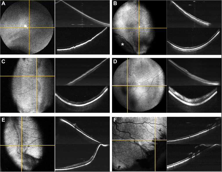
Acquisition of and B-scan imaging of the peripheral retina to objectively assess early vitreoretinal pathology, including the demarcation between vascularized and anterior avascular retina, the presence of early ridge formation, and small neovascular tufts.

RESULTS

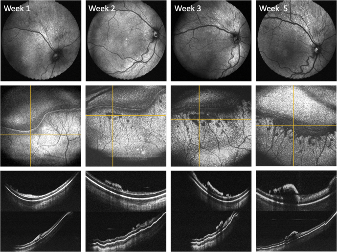
Various stages of ROP were detected using a rapid acquisition OCT system. In one neonate, serial OCT imaging over a five-week period demonstrated accumulation of neovascular tufts with progression to stage 3 ROP with extraretinal fibrovascular proliferation along the ridge. Videography of this technique is included in this report for instructional purposes.

CONCLUSIONS

Serial examinations using widefield OCT and scleral depression is feasible and may improve detection and documentation of ROP disease progression. Earlier detection of ROP-related proliferation may prevent vitreoretinal traction, retinal detachment, and blindness.

摘要

目的

确定在使用巩膜压迫法改善周边视野时,手持宽视野光学相干断层扫描(OCT)是否可用于记录早产儿视网膜病变(ROP)分期。

设计

前瞻性观察性研究。

参与者

在单一学术医疗中心新生儿重症监护病房(NICU)收治的连续新生儿,这些新生儿也符合ROP筛查标准并同意进行研究成像。

方法

在2020年10月28日至2021年3月3日的常规ROP筛查期间,使用研究性的400千赫、55度视野手持OCT将巩膜压迫法与宽视野OCT相结合。

主要观察指标

获取周边视网膜的A扫描和B扫描成像,以客观评估早期玻璃体视网膜病变,包括血管化视网膜与前部无血管视网膜之间的界限、早期嵴形成的存在以及小的新生血管簇。

结果

使用快速采集OCT系统检测到了不同阶段的ROP。在一名新生儿中,为期五周的连续OCT成像显示新生血管簇积聚,并进展为3期ROP,沿嵴出现视网膜外纤维血管增殖。本报告包含该技术的视频记录以供教学使用。

结论

使用宽视野OCT和巩膜压迫法进行连续检查是可行的,可能会改善ROP疾病进展的检测和记录。早期检测ROP相关增殖可能预防玻璃体视网膜牵拉、视网膜脱离和失明。

https://cdn.ncbi.nlm.nih.gov/pmc/blobs/1f03/9559887/5dde771eccff/gr2.jpg
https://cdn.ncbi.nlm.nih.gov/pmc/blobs/1f03/9559887/baaa8ebc75fa/gr1.jpg
https://cdn.ncbi.nlm.nih.gov/pmc/blobs/1f03/9559887/5dde771eccff/gr2.jpg
https://cdn.ncbi.nlm.nih.gov/pmc/blobs/1f03/9559887/baaa8ebc75fa/gr1.jpg
https://cdn.ncbi.nlm.nih.gov/pmc/blobs/1f03/9559887/5dde771eccff/gr2.jpg

相似文献

1
Peripheral OCT Assisted by Scleral Depression in Retinopathy of Prematurity.巩膜压迫辅助下的周边光学相干断层扫描在早产儿视网膜病变中的应用
Ophthalmol Sci. 2022 Mar;2(1). doi: 10.1016/j.xops.2021.100094. Epub 2022 Jan 11.
2
Handheld Optical Coherence Tomography Angiography and Ultra-Wide-Field Optical Coherence Tomography in Retinopathy of Prematurity.手持式光学相干断层扫描血管造影术和超广角光学相干断层扫描在早产儿视网膜病变中的应用
JAMA Ophthalmol. 2017 Sep 1;135(9):977-981. doi: 10.1001/jamaophthalmol.2017.2481.
3
Integrated Visualization Highlighting Retinal Changes in Retinopathy of Prematurity From 3-Dimensional Optical Coherence Tomography Data.基于三维光学相干断层扫描数据的综合可视化突出早产儿视网膜病变的视网膜变化。
JAMA Ophthalmol. 2022 Jul 1;140(7):725-729. doi: 10.1001/jamaophthalmol.2022.1344.
4
Association of Optical Coherence Tomography-Measured Fibrovascular Ridge Thickness and Clinical Disease Stage in Retinopathy of Prematurity.光学相干断层扫描测量的纤维血管嵴厚度与早产儿视网膜病变临床疾病分期的相关性
JAMA Ophthalmol. 2022 Oct 13;140(11):1121-7. doi: 10.1001/jamaophthalmol.2022.4173.
5
Differentiating Retinal Detachment and Retinoschisis Using Handheld Optical Coherence Tomography in Stage 4 Retinopathy of Prematurity.手持式光学相干断层扫描在 4 期早产儿视网膜病变中对视网膜脱离和视网膜劈裂的鉴别诊断。
JAMA Ophthalmol. 2020 Jan 1;138(1):81-85. doi: 10.1001/jamaophthalmol.2019.4796.
6
Three-dimensional pattern of extraretinal neovascular development in retinopathy of prematurity.早产儿视网膜病变中视网膜外新生血管发育的三维模式
Graefes Arch Clin Exp Ophthalmol. 2019 Apr;257(4):677-688. doi: 10.1007/s00417-019-04274-6. Epub 2019 Feb 21.
7
Spectral-Domain OCT Findings of Retinal Vascular-Avascular Junction in Infants with Retinopathy of Prematurity.早产儿视网膜病变婴儿视网膜血管-无血管交界处的频域光学相干断层扫描结果
Ophthalmol Retina. 2018 Sep;2(9):963-971. doi: 10.1016/j.oret.2018.02.001. Epub 2018 Mar 21.
8
Retinoschisis detected with handheld spectral-domain optical coherence tomography in neonates with advanced retinopathy of prematurity.在患有晚期早产儿视网膜病变的新生儿中,通过手持式光谱域光学相干断层扫描检测到视网膜劈裂。
Arch Ophthalmol. 2010 Jan;128(1):57-62. doi: 10.1001/archophthalmol.2009.361.
9
Advantages of Widefield Optical Coherence Tomography in the Diagnosis of Retinopathy of Prematurity.广角光学相干断层扫描在早产儿视网膜病变诊断中的优势
Front Pediatr. 2022 Jan 18;9:797684. doi: 10.3389/fped.2021.797684. eCollection 2021.
10
Insights into advanced retinopathy of prematurity using handheld spectral domain optical coherence tomography imaging.利用手持式光谱域光学相干断层扫描成像技术对早产儿晚期视网膜病变的深入研究
Ophthalmology. 2009 Dec;116(12):2448-56. doi: 10.1016/j.ophtha.2009.06.003. Epub 2009 Sep 18.

引用本文的文献

1
Ultra-Widefield Optical Coherence Tomography Beyond the Ora Serrata in Retinopathy of Prematurity.早产儿视网膜病变中超越锯齿缘的超广角光学相干断层扫描
JAMA Ophthalmol. 2025 Feb 1;143(2):165-170. doi: 10.1001/jamaophthalmol.2024.5533.
2
Panretinal handheld OCT angiography for pediatric retinal imaging.用于儿科视网膜成像的全视网膜手持式光学相干断层扫描血管造影术。
Biomed Opt Express. 2024 Apr 29;15(5):3412-3424. doi: 10.1364/BOE.520739. eCollection 2024 May 1.
3
Implementation of optical coherence tomography in retinopathy of prematurity screening.

本文引用的文献

1
High-speed and widefield handheld swept-source OCT angiography with a VCSEL light source.采用垂直腔面发射激光器(VCSEL)光源的高速宽视野手持式扫频源光学相干断层扫描血管造影术。
Biomed Opt Express. 2021 May 20;12(6):3553-3570. doi: 10.1364/BOE.425411. eCollection 2021 Jun 1.
2
Preterm Infant Stress During Handheld Optical Coherence Tomography vs Binocular Indirect Ophthalmoscopy Examination for Retinopathy of Prematurity.早产儿手持光学相干断层扫描与双目间接检眼镜检查早产儿视网膜病变时的应激反应。
JAMA Ophthalmol. 2021 May 1;139(5):567-574. doi: 10.1001/jamaophthalmol.2021.0377.
3
Evaluation of a Deep Learning-Derived Quantitative Retinopathy of Prematurity Severity Scale.
在早产儿视网膜病变筛查中实施光学相干断层扫描。
Curr Opin Ophthalmol. 2024 May 1;35(3):252-259. doi: 10.1097/ICU.0000000000001030. Epub 2024 Jan 11.
4
Vitreoretinal biomarkers of retinopathy of prematurity using handheld optical coherence tomography: a review.使用手持式光学相干断层扫描技术检测早产儿视网膜病变的玻璃体视网膜生物标志物:综述
Front Pediatr. 2023 May 31;11:1191174. doi: 10.3389/fped.2023.1191174. eCollection 2023.
5
Multivariate Models to Diagnose Early Referral-Warranted Retinopathy of Prematurity With Handheld Optical Coherence Tomography.应用手持式光学相干断层扫描仪对早产儿视网膜病变进行早期转诊相关诊断的多元模型分析。
Transl Vis Sci Technol. 2023 May 1;12(5):26. doi: 10.1167/tvst.12.5.26.
6
Panretinal Optical Coherence Tomography.全视网膜光学相干断层扫描。
IEEE Trans Med Imaging. 2023 Nov;42(11):3219-3228. doi: 10.1109/TMI.2023.3278269. Epub 2023 Oct 27.
7
Latest Trends in Retinopathy of Prematurity: Research on Risk Factors, Diagnostic Methods and Therapies.早产儿视网膜病变的最新趋势:危险因素、诊断方法及治疗的研究
Int J Gen Med. 2023 Mar 14;16:937-949. doi: 10.2147/IJGM.S401122. eCollection 2023.
8
Association of Optical Coherence Tomography-Measured Fibrovascular Ridge Thickness and Clinical Disease Stage in Retinopathy of Prematurity.光学相干断层扫描测量的纤维血管嵴厚度与早产儿视网膜病变临床疾病分期的相关性
JAMA Ophthalmol. 2022 Oct 13;140(11):1121-7. doi: 10.1001/jamaophthalmol.2022.4173.
9
Widefield Optical Coherence Tomography in Pediatric Retina: A Case Series of Intraoperative Applications Using a Prototype Handheld Device.小儿视网膜广角光学相干断层扫描:使用原型手持设备的术中应用病例系列
Front Med (Lausanne). 2022 Jul 4;9:860371. doi: 10.3389/fmed.2022.860371. eCollection 2022.
10
Insights into the developing fovea revealed by imaging.成像技术揭示的黄斑发育奥秘。
Prog Retin Eye Res. 2022 Sep;90:101067. doi: 10.1016/j.preteyeres.2022.101067. Epub 2022 May 17.
深度学习定量早产儿视网膜病变严重程度评分评估。
Ophthalmology. 2021 Jul;128(7):1070-1076. doi: 10.1016/j.ophtha.2020.10.025. Epub 2020 Oct 27.
4
Macular OCT Characteristics at 36 Weeks' Postmenstrual Age in Infants Examined for Retinopathy of Prematurity.早产儿视网膜病变筛查婴儿在 36 孕周时的黄斑 OCT 特征。
Ophthalmol Retina. 2021 Jun;5(6):580-592. doi: 10.1016/j.oret.2020.09.004. Epub 2020 Sep 11.
5
Vitreous Findings by Handheld Spectral-Domain OCT Correlate with Retinopathy of Prematurity Severity.手持式光谱域光学相干断层扫描的玻璃体检查结果与早产儿视网膜病变严重程度相关。
Ophthalmol Retina. 2020 Oct;4(10):1008-1015. doi: 10.1016/j.oret.2020.03.027. Epub 2020 Apr 9.
6
Spectral-Domain OCT Findings of Retinal Vascular-Avascular Junction in Infants with Retinopathy of Prematurity.早产儿视网膜病变婴儿视网膜血管-无血管交界处的频域光学相干断层扫描结果
Ophthalmol Retina. 2018 Sep;2(9):963-971. doi: 10.1016/j.oret.2018.02.001. Epub 2018 Mar 21.
7
Handheld Optical Coherence Tomography Angiography and Ultra-Wide-Field Optical Coherence Tomography in Retinopathy of Prematurity.手持式光学相干断层扫描血管造影术和超广角光学相干断层扫描在早产儿视网膜病变中的应用
JAMA Ophthalmol. 2017 Sep 1;135(9):977-981. doi: 10.1001/jamaophthalmol.2017.2481.
8
Retinal Imaging of Infants on Spectral Domain Optical Coherence Tomography.婴儿视网膜的光谱域光学相干断层扫描成像
Biomed Res Int. 2015;2015:782420. doi: 10.1155/2015/782420. Epub 2015 Jul 6.
9
Poorer neurodevelopmental outcomes associated with cystoid macular edema identified in preterm infants in the intensive care nursery.在重症监护病房的早产儿中,发现与黄斑囊样水肿相关的神经发育结局较差。
Ophthalmology. 2015 Mar;122(3):610-9. doi: 10.1016/j.ophtha.2014.09.022. Epub 2014 Nov 4.
10
Three-dimensional assessment of vascular and perivascular characteristics in subjects with retinopathy of prematurity.早产儿视网膜病变患者血管和血管周围特征的三维评估。
Ophthalmology. 2014 Jun;121(6):1289-96. doi: 10.1016/j.ophtha.2013.12.004. Epub 2014 Jan 21.