Department of Gastroenterology, West China Hospital, Sichuan University, Chengdu, Sichuan, China.
Department of Gastroenterology, Huai'an First People's Hospital, Huai'an, Jiangsu, China.
Clin Transl Gastroenterol. 2022 Jan 11;13(1):e00433. doi: 10.14309/ctg.0000000000000433.
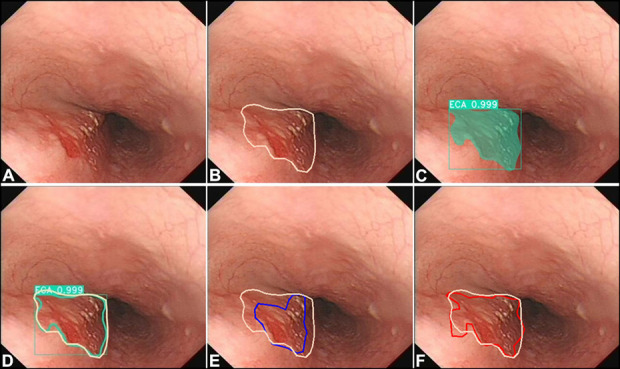
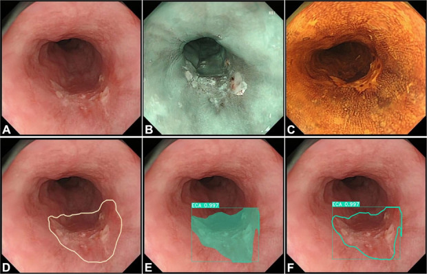
Conventional white light imaging (WLI) endoscopy is the most common screening technique used for detecting early esophageal squamous cell carcinoma (ESCC). Nevertheless, it is difficult to detect and delineate margins of early ESCC using WLI endoscopy. This study aimed to develop an artificial intelligence (AI) model to detect and delineate margins of early ESCC under WLI endoscopy.
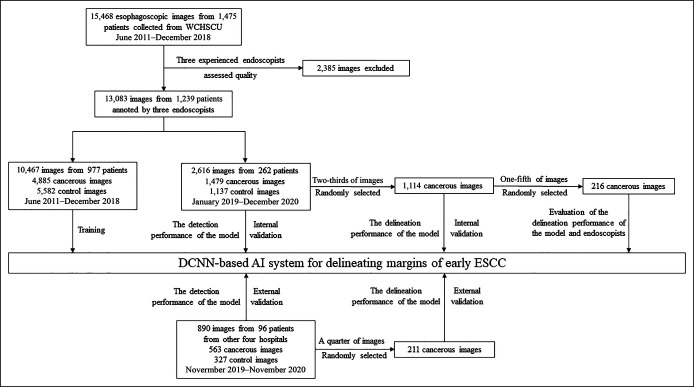
A total of 13,083 WLI images from 1,239 patients were used to train and test the AI model. To evaluate the detection performance of the model, 1,479 images and 563 images were used as internal and external validation data sets, respectively. For assessing the delineation performance of the model, 1,114 images and 211 images were used as internal and external validation data sets, respectively. In addition, 216 images were used to compare the delineation performance between the model and endoscopists.
The model showed an accuracy of 85.7% and 84.5% in detecting lesions in internal and external validation, respectively. For delineating margins, the model achieved an accuracy of 93.4% and 95.7% in the internal and external validation, respectively, under an overlap ratio of 0.60. The accuracy of the model, senior endoscopists, and expert endoscopists in delineating margins were 98.1%, 78.6%, and 95.3%, respectively. The proposed model achieved similar delineating performance compared with that of expert endoscopists but superior to senior endoscopists.
We successfully developed an AI model, which can be used to accurately detect early ESCC and delineate the margins of the lesions under WLI endoscopy.
传统的白光成像(WLI)内镜检查是最常用的筛查技术,用于检测早期食管鳞状细胞癌(ESCC)。然而,使用 WLI 内镜检查很难检测和描绘早期 ESCC 的边界。本研究旨在开发一种人工智能(AI)模型,用于检测和描绘 WLI 内镜下早期 ESCC 的边界。
共使用了 1239 名患者的 13083 张 WLI 图像来训练和测试 AI 模型。为了评估模型的检测性能,分别使用了 1479 张和 563 张图像作为内部和外部验证数据集。为了评估模型的描绘性能,分别使用了 1114 张和 211 张图像作为内部和外部验证数据集。此外,还使用了 216 张图像来比较模型和内镜医生的描绘性能。
该模型在内部和外部验证中的检测病变的准确率分别为 85.7%和 84.5%。对于边界描绘,该模型在重叠比为 0.60 时,在内部和外部验证中的准确率分别为 93.4%和 95.7%。模型、高级内镜医生和专家内镜医生在边界描绘方面的准确率分别为 98.1%、78.6%和 95.3%。与专家内镜医生相比,该模型具有相似的描绘性能,但优于高级内镜医生。
我们成功开发了一种人工智能模型,可用于准确检测 WLI 内镜下的早期 ESCC 并描绘病变的边界。