• 文献检索
  • 文档翻译
  • 深度研究
  • 学术资讯
  • Suppr Zotero 插件Zotero 插件
  • 邀请有礼
  • 套餐&价格
  • 历史记录
应用&插件
Suppr Zotero 插件Zotero 插件浏览器插件Mac 客户端Windows 客户端微信小程序
定价
高级版会员购买积分包购买API积分包
服务
文献检索文档翻译深度研究API 文档MCP 服务
关于我们
关于 Suppr公司介绍联系我们用户协议隐私条款
关注我们

Suppr 超能文献

核心技术专利:CN118964589B侵权必究
粤ICP备2023148730 号-1Suppr @ 2026

文献检索

告别复杂PubMed语法,用中文像聊天一样搜索,搜遍4000万医学文献。AI智能推荐,让科研检索更轻松。

立即免费搜索

文件翻译

保留排版,准确专业,支持PDF/Word/PPT等文件格式,支持 12+语言互译。

免费翻译文档

深度研究

AI帮你快速写综述,25分钟生成高质量综述,智能提取关键信息,辅助科研写作。

立即免费体验

乳腺成纤维细胞的免疫调节及乳腺X线密度的影响

Immune Regulation of Mammary Fibroblasts and the Impact of Mammographic Density.

作者信息

Archer Maddison, Dasari Pallave, Walsh David, Britt Kara L, Evdokiou Andreas, Ingman Wendy V

机构信息

Discipline of Surgical Specialties, Adelaide Medical School, The Queen Elizabeth Hospital, University of Adelaide, Adelaide, SA 5011, Australia.

Robinson Research Institute, University of Adelaide, Adelaide, SA 5001, Australia.

出版信息

J Clin Med. 2022 Feb 2;11(3):799. doi: 10.3390/jcm11030799.

DOI:10.3390/jcm11030799
PMID:35160252
原文链接:https://pmc.ncbi.nlm.nih.gov/articles/PMC8837019/
Abstract

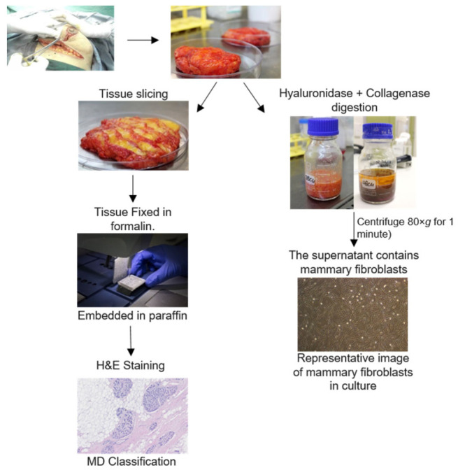
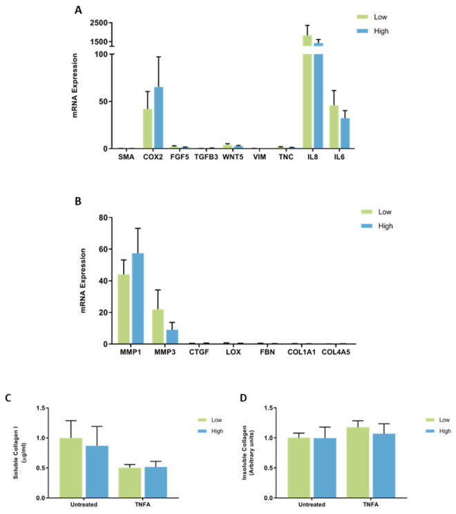
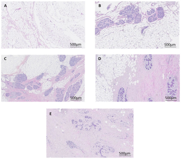
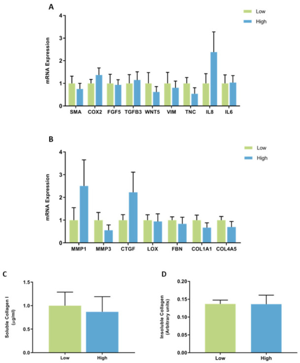
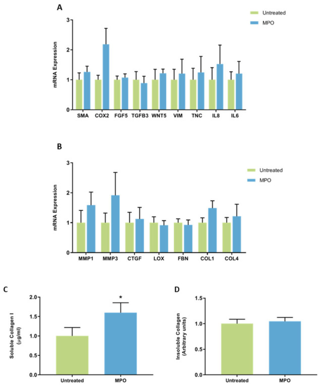
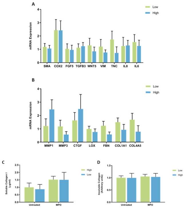
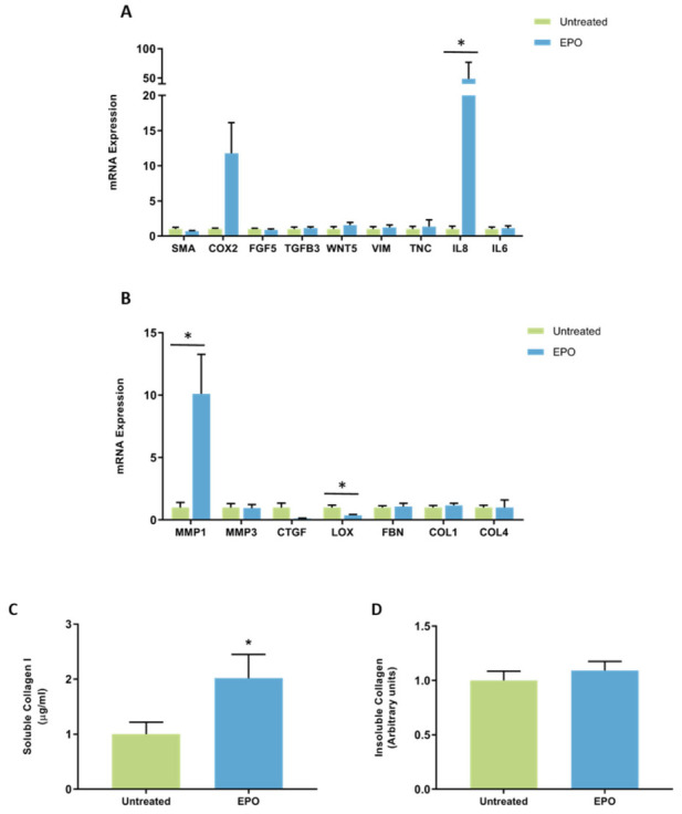
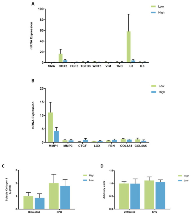
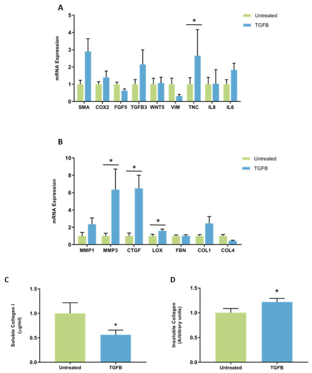
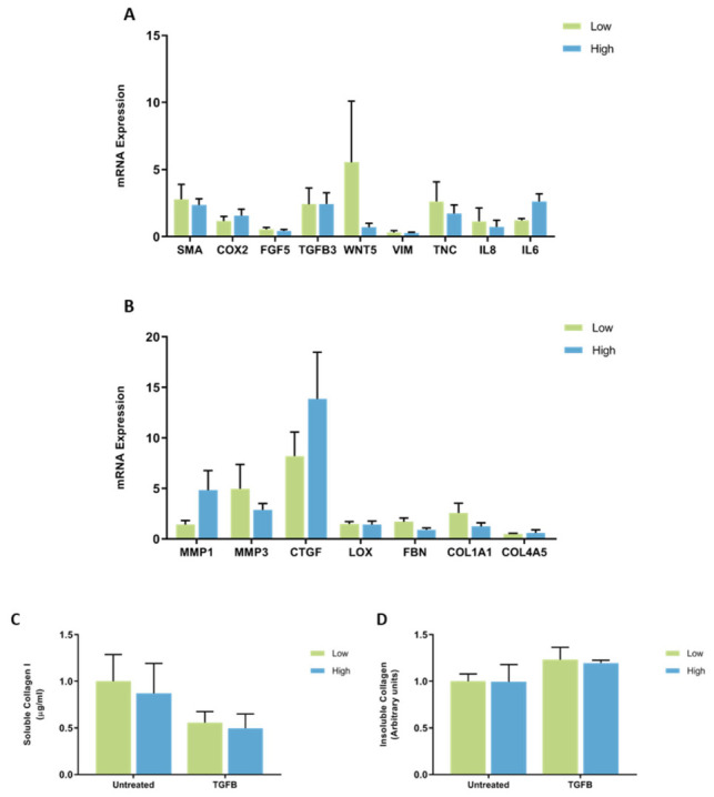
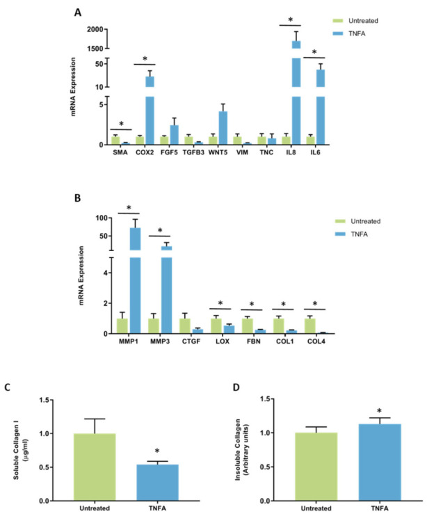
Mammographic density is associated with a 4-6-fold increase in breast cancer risk independent of age and BMI. High mammographic density is characterized by breast tissue with high proportions of stroma comprised of fibroblasts, collagen, and immune cells. This study sought to investigate whether stromal fibroblasts from high mammographic density breast tissue contributes to increased extracellular matrix deposition and pro-tumorigenic signaling. Mammary fibroblasts were isolated from women with high and low mammographic density and exposed to immune factors myeloperoxidase (MPO), eosinophil peroxidase (EPO), transforming growth factor beta 1 (TGFB1) and tumour necrosis factor alpha (TNFA) for 72 h and profiled for expression of cancer-associated fibroblast and extracellular matrix regulation markers. No differences in gene expression profiles or collagen production were observed between fibroblasts with high or low mammographic density, and they did not have a differential response to immune mediators. MPO and EPO significantly increased the production of collagen 1. TGFB and TNFA induced variable changes in gene expression. Fibroblasts cultured in vitro from women with high mammographic density do not appear to be inherently different to those from women with low mammographic density. The function of fibroblasts in mammographic density-associated breast cancer risk is likely to be regulated by immune signals from surrounding cells in the microenvironment.

摘要

乳腺钼靶密度与乳腺癌风险增加4至6倍相关,且独立于年龄和体重指数。高乳腺钼靶密度的特征是乳腺组织中由成纤维细胞、胶原蛋白和免疫细胞组成的基质比例较高。本研究旨在调查来自高乳腺钼靶密度乳腺组织的基质成纤维细胞是否会导致细胞外基质沉积增加和促肿瘤信号传导。从乳腺钼靶密度高和低的女性中分离出乳腺成纤维细胞,并使其暴露于免疫因子髓过氧化物酶(MPO)、嗜酸性粒细胞过氧化物酶(EPO)、转化生长因子β1(TGFB1)和肿瘤坏死因子α(TNFA)72小时,并分析癌症相关成纤维细胞和细胞外基质调节标志物的表达。在乳腺钼靶密度高或低的成纤维细胞之间未观察到基因表达谱或胶原蛋白产生的差异,并且它们对免疫介质没有差异反应。MPO和EPO显著增加了胶原蛋白1的产生。TGFB和TNFA诱导了基因表达的可变变化。从乳腺钼靶密度高的女性体外培养的成纤维细胞似乎与乳腺钼靶密度低的女性的成纤维细胞没有本质区别。成纤维细胞在乳腺钼靶密度相关乳腺癌风险中的功能可能受微环境中周围细胞的免疫信号调节。

https://cdn.ncbi.nlm.nih.gov/pmc/blobs/fc02/8837019/4b3abfd66855/jcm-11-00799-g011.jpg
https://cdn.ncbi.nlm.nih.gov/pmc/blobs/fc02/8837019/69bf3b1a9d41/jcm-11-00799-g001.jpg
https://cdn.ncbi.nlm.nih.gov/pmc/blobs/fc02/8837019/dcb3a903024e/jcm-11-00799-g002.jpg
https://cdn.ncbi.nlm.nih.gov/pmc/blobs/fc02/8837019/7c9d73cc6ee9/jcm-11-00799-g003.jpg
https://cdn.ncbi.nlm.nih.gov/pmc/blobs/fc02/8837019/16178df3d3fc/jcm-11-00799-g004.jpg
https://cdn.ncbi.nlm.nih.gov/pmc/blobs/fc02/8837019/3fe76aa61a1b/jcm-11-00799-g005.jpg
https://cdn.ncbi.nlm.nih.gov/pmc/blobs/fc02/8837019/497e92ad00f2/jcm-11-00799-g006.jpg
https://cdn.ncbi.nlm.nih.gov/pmc/blobs/fc02/8837019/e971aa68c1e0/jcm-11-00799-g007.jpg
https://cdn.ncbi.nlm.nih.gov/pmc/blobs/fc02/8837019/1024653256f4/jcm-11-00799-g008.jpg
https://cdn.ncbi.nlm.nih.gov/pmc/blobs/fc02/8837019/a207189b4fb5/jcm-11-00799-g009.jpg
https://cdn.ncbi.nlm.nih.gov/pmc/blobs/fc02/8837019/0411dd9a80f8/jcm-11-00799-g010.jpg
https://cdn.ncbi.nlm.nih.gov/pmc/blobs/fc02/8837019/4b3abfd66855/jcm-11-00799-g011.jpg

相似文献

1
Immune Regulation of Mammary Fibroblasts and the Impact of Mammographic Density.乳腺成纤维细胞的免疫调节及乳腺X线密度的影响
J Clin Med. 2022 Feb 2;11(3):799. doi: 10.3390/jcm11030799.
2
Biological Mechanisms and Therapeutic Opportunities in Mammographic Density and Breast Cancer Risk.乳腺X线密度与乳腺癌风险的生物学机制及治疗机遇
Cancers (Basel). 2021 Oct 27;13(21):5391. doi: 10.3390/cancers13215391.
3
Inflammatory peroxidases promote breast cancer progression in mice via regulation of the tumour microenvironment.炎症性过氧化物酶通过调节肿瘤微环境促进小鼠乳腺癌进展。
Int J Oncol. 2017 Apr;50(4):1191-1200. doi: 10.3892/ijo.2017.3883. Epub 2017 Feb 20.
4
High mammographic density is associated with an increase in stromal collagen and immune cells within the mammary epithelium.乳腺钼靶高密度与乳腺上皮内基质胶原蛋白和免疫细胞增加有关。
Breast Cancer Res. 2015 Jun 4;17(1):79. doi: 10.1186/s13058-015-0592-1.
5
JNK1 stress signaling is hyper-activated in high breast density and the tumor stroma: connecting fibrosis, inflammation, and stemness for cancer prevention.JNK1应激信号在乳腺密度高和肿瘤基质中过度激活:将纤维化、炎症和干性联系起来以预防癌症。
Cell Cycle. 2014;13(4):580-99. doi: 10.4161/cc.27379. Epub 2013 Dec 5.
6
Pubertal mammary gland development is a key determinant of adult mammographic density.青春期乳腺发育是成年乳腺 X 光密度的关键决定因素。
Semin Cell Dev Biol. 2021 Jun;114:143-158. doi: 10.1016/j.semcdb.2020.11.011. Epub 2020 Dec 11.
7
Decreased TGFbeta signaling and increased COX2 expression in high risk women with increased mammographic breast density.乳腺钼靶密度增加的高危女性中转化生长因子β信号传导降低且环氧化酶2表达增加。
Breast Cancer Res Treat. 2010 Jan;119(2):305-14. doi: 10.1007/s10549-009-0350-0. Epub 2009 Feb 25.
8
Stress signaling from human mammary epithelial cells contributes to phenotypes of mammographic density.来自人乳腺上皮细胞的应激信号传导有助于乳房X线密度的表型。
Cancer Res. 2014 Sep 15;74(18):5032-5044. doi: 10.1158/0008-5472.CAN-13-3390. Epub 2014 Aug 29.
9
Amount of stroma is associated with mammographic density and stromal expression of oestrogen receptor in normal breast tissues.在正常乳腺组织中,间质数量与乳腺X线密度以及雌激素受体的间质表达相关。
Breast Cancer Res Treat. 2016 Jul;158(2):253-61. doi: 10.1007/s10549-016-3877-x. Epub 2016 Jun 27.
10
Activation of Transforming Growth Factor Beta 1 Signaling in Gastric Cancer-associated Fibroblasts Increases Their Motility, via Expression of Rhomboid 5 Homolog 2, and Ability to Induce Invasiveness of Gastric Cancer Cells.胃癌相关成纤维细胞中转化生长因子β 1 信号的激活通过表达菱形 5 同源物 2 增加其运动性,并增强诱导胃癌细胞侵袭的能力。
Gastroenterology. 2017 Jul;153(1):191-204.e16. doi: 10.1053/j.gastro.2017.03.046. Epub 2017 Apr 5.

引用本文的文献

1
A Deep Learning Approach for the Classification of Fibroglandular Breast Density in Histology Images of Human Breast Tissue.一种用于人类乳腺组织组织学图像中纤维腺体型乳腺密度分类的深度学习方法。
Cancers (Basel). 2025 Jan 28;17(3):449. doi: 10.3390/cancers17030449.
2
Association between breastfeeding, mammographic density, and breast cancer risk: a review.母乳喂养、乳腺密度与乳腺癌风险的关系:综述。
Int Breastfeed J. 2024 Sep 16;19(1):65. doi: 10.1186/s13006-024-00672-7.
3
CCL2-Mediated Stromal Interactions Drive Macrophage Polarization to Increase Breast Tumorigenesis.

本文引用的文献

1
Aging-Associated Alterations in Mammary Epithelia and Stroma Revealed by Single-Cell RNA Sequencing.单细胞 RNA 测序揭示的与衰老相关的乳腺上皮和基质改变。
Cell Rep. 2020 Dec 29;33(13):108566. doi: 10.1016/j.celrep.2020.108566.
2
Mitochondrial mutations and mitoepigenetics: Focus on regulation of oxidative stress-induced responses in breast cancers.线粒体突变与线粒体表观遗传学:聚焦于调控乳腺癌中氧化应激诱导反应。
Semin Cancer Biol. 2022 Aug;83:556-569. doi: 10.1016/j.semcancer.2020.09.012. Epub 2020 Oct 6.
3
Lysyl oxidase enzymes mediate TGF-β1-induced fibrotic phenotypes in human skin-like tissues.
CCL2 介导体细胞相互作用驱动巨噬细胞极化以增加乳腺癌发生。
Int J Mol Sci. 2023 Apr 17;24(8):7385. doi: 10.3390/ijms24087385.
4
Estrogen-induced immune changes within the normal mammary gland.雌激素诱导的正常乳腺内免疫变化。
Sci Rep. 2022 Nov 8;12(1):18986. doi: 10.1038/s41598-022-21871-4.
赖氨酰氧化酶酶介导 TGF-β1 在人皮肤样组织中诱导的纤维化表型。
Lab Invest. 2019 Apr;99(4):514-527. doi: 10.1038/s41374-018-0159-8. Epub 2018 Dec 19.
4
Cancer-associated fibroblasts as key regulators of the breast cancer tumor microenvironment.癌症相关成纤维细胞作为乳腺癌肿瘤微环境的关键调节者。
Cancer Metastasis Rev. 2018 Dec;37(4):577-597. doi: 10.1007/s10555-018-9768-3.
5
High mammographic density in women is associated with protumor inflammation.女性乳腺密度高与肿瘤前炎症有关。
Breast Cancer Res. 2018 Aug 9;20(1):92. doi: 10.1186/s13058-018-1010-2.
6
Volumetric Mammographic Density, Age-Related Decline, and Breast Cancer Risk Factors in a National Breast Cancer Screening Program.容积乳腺密度、与年龄相关的下降以及国家乳腺癌筛查计划中的乳腺癌危险因素。
Cancer Epidemiol Biomarkers Prev. 2018 Sep;27(9):1065-1074. doi: 10.1158/1055-9965.EPI-18-0151. Epub 2018 Jun 20.
7
A peek into cancer-associated fibroblasts: origins, functions and translational impact.浅析癌症相关成纤维细胞:起源、功能及转化影响。
Dis Model Mech. 2018 Apr 19;11(4):dmm029447. doi: 10.1242/dmm.029447.
8
Myeloperoxidase as an Active Disease Biomarker: Recent Biochemical and Pathological Perspectives.髓过氧化物酶作为一种活性疾病生物标志物:最新的生化与病理学观点
Med Sci (Basel). 2018 Apr 18;6(2):33. doi: 10.3390/medsci6020033.
9
Lysyl Oxidase: Its Diversity in Health and Diseases.赖氨酰氧化酶:其在健康与疾病中的多样性
Indian J Clin Biochem. 2017 Jun;32(2):134-141. doi: 10.1007/s12291-016-0576-7. Epub 2016 May 11.
10
Comparative analysis of lysyl oxidase (like) family members in pulmonary fibrosis.肺纤维化中赖氨酰氧化酶(类似)家族成员的比较分析。
Sci Rep. 2017 Mar 10;7(1):149. doi: 10.1038/s41598-017-00270-0.