Toure Abdoulaye, Gnaoulé Debato Tina, Judicael Ahoury, Ndja Ange Patrick, Zouzou Ange Eric, Dion Anicet Le, Fatto Nguessan Ebeyss, Gbazi Gogoua Casimir
Radiology Department, University Hospital of Cocody, 25 P.O. Box 2168 Abidjan 25, Abidjan, Cote d'Ivoire.
School of Medicine, Félix Houphouët Boigny University, Abidjan, Cote d'Ivoire.
Radiol Case Rep. 2022 Feb 3;17(4):1068-1075. doi: 10.1016/j.radcr.2022.01.049. eCollection 2022 Apr.
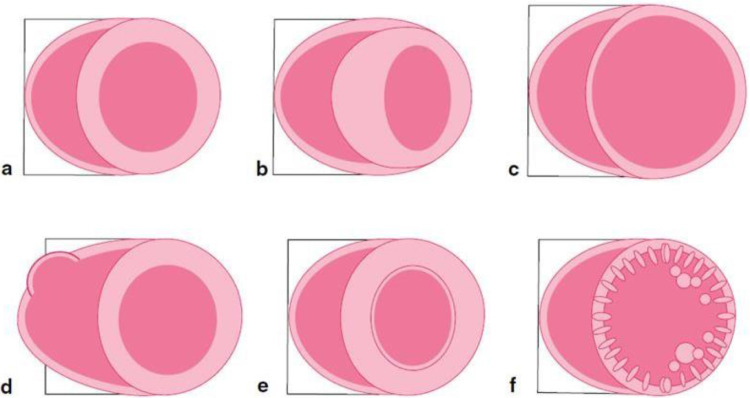
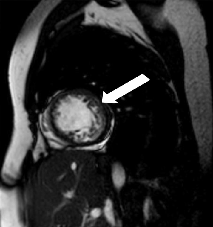
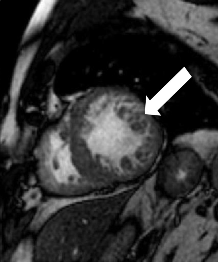
Left ventricular non compaction (LVNC) is a relatively rare variety of cardiomyopathy of genetic origin. We report three cases of LVNC diagnosed on cardiac magnetic resonance imaging (MRI) in Abidjan in patients aged 42, 46 and 60 years, referred for suspected LVNC on echocardiography. We used a 1.5 T MRI and performed the following sequences: black blood and white blood, LV minor axis, LV major axis, 4 cavities, and T1 SPIR Gadolinium (early and late enhancement at 10 minutes). MRI made the diagnosis of LVNC based on a double-layered myocardium, the inner (endocardium) non compacted, fibrillar thickened and the outer (epicardium) compacted thin with a non compacted to compacted myocardium ratio greater than 2.3, making the formal diagnosis. Cardiac MRI is an excellent diagnostic tool for LVNC. Its recent use in Africa should be common in the management of this cardiomyopathy.
左心室心肌致密化不全(LVNC)是一种相对罕见的遗传性心肌病。我们报告了在阿比让通过心脏磁共振成像(MRI)诊断出的3例LVNC病例,患者年龄分别为42岁、46岁和60岁,因超声心动图怀疑为LVNC而转诊。我们使用1.5T MRI并进行了以下序列检查:黑血和白血序列、左心室短轴、左心室长轴、四腔心以及T1脂肪抑制钆增强序列(10分钟时的早期和晚期强化)。MRI基于双层心肌做出LVNC的诊断,内层(心内膜)心肌未致密化、纤维增厚,外层(心外膜)致密化且薄,未致密化心肌与致密化心肌的比例大于2.3,从而做出明确诊断。心脏MRI是诊断LVNC的优秀工具。其近期在非洲的应用在这种心肌病的管理中应该会很常见。