• 文献检索
  • 文档翻译
  • 深度研究
  • 学术资讯
  • Suppr Zotero 插件Zotero 插件
  • 邀请有礼
  • 套餐&价格
  • 历史记录
应用&插件
Suppr Zotero 插件Zotero 插件浏览器插件Mac 客户端Windows 客户端微信小程序
定价
高级版会员购买积分包购买API积分包
服务
文献检索文档翻译深度研究API 文档MCP 服务
关于我们
关于 Suppr公司介绍联系我们用户协议隐私条款
关注我们

Suppr 超能文献

核心技术专利:CN118964589B侵权必究
粤ICP备2023148730 号-1Suppr @ 2026

文献检索

告别复杂PubMed语法,用中文像聊天一样搜索,搜遍4000万医学文献。AI智能推荐,让科研检索更轻松。

立即免费搜索

文件翻译

保留排版,准确专业,支持PDF/Word/PPT等文件格式,支持 12+语言互译。

免费翻译文档

深度研究

AI帮你快速写综述,25分钟生成高质量综述,智能提取关键信息,辅助科研写作。

立即免费体验

通过无监督3D细胞核分割分析细胞-支架相互作用

Analyzing Cell-Scaffold Interaction through Unsupervised 3D Nuclei Segmentation.

作者信息

Yao Kai, Sun Jie, Huang Kaizhu, Jing Linzhi, Liu Hang, Huang Dejian, Jude Curran

机构信息

School of Advanced Technology, Xi'an Jiaotong-Liverpool University, 111 Ren'ai Road, Suzhou, Jiangsu 215123, China.

School of Engineering, University of Liverpool, The Quadrangle, Brownlow Hill, L69 3GH, UK.

出版信息

Int J Bioprint. 2021 Dec 30;8(1):495. doi: 10.18063/ijb.v8i1.495. eCollection 2022.

DOI:10.18063/ijb.v8i1.495
PMID:35187282
原文链接:https://pmc.ncbi.nlm.nih.gov/articles/PMC8852265/
Abstract

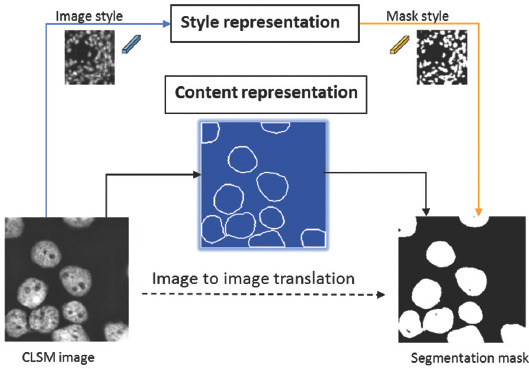
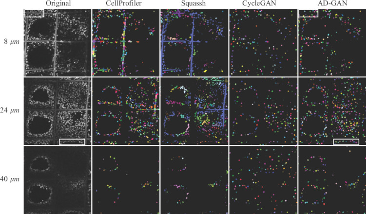
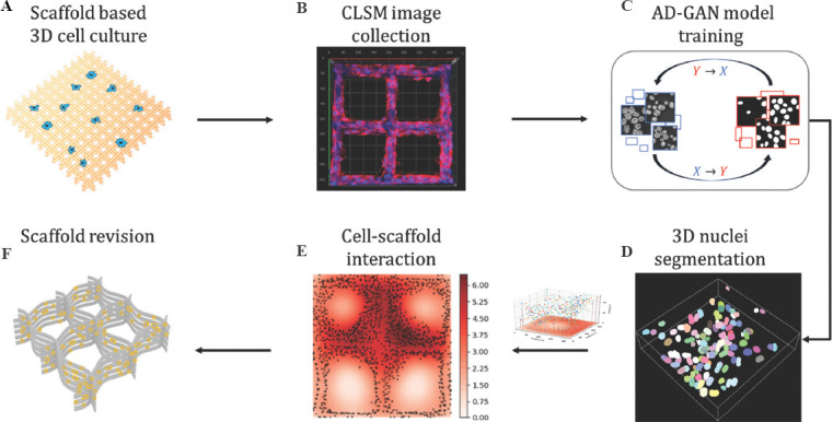
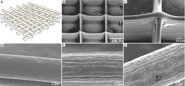
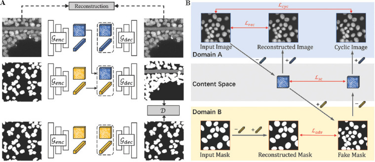
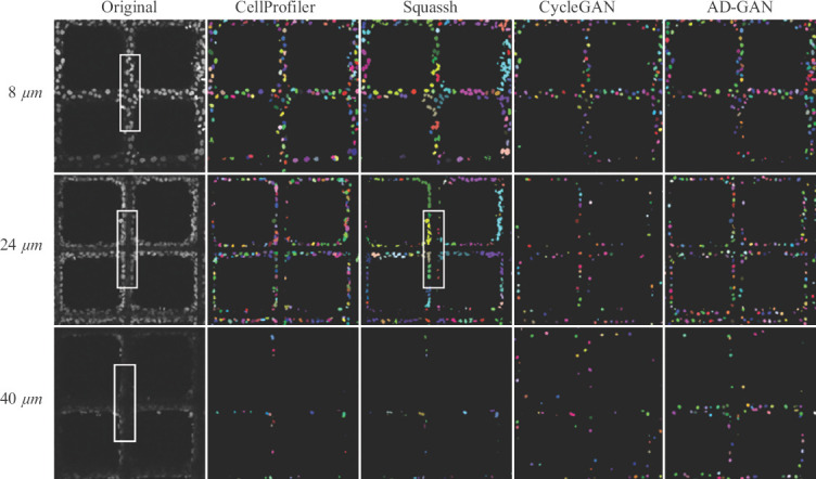
Fibrous scaffolds have been extensively used in three-dimensional (3D) cell culture systems to establish models in cell biology, tissue engineering, and drug screening. It is a common practice to characterize cell behaviors on such scaffolds using confocal laser scanning microscopy (CLSM). As a noninvasive technology, CLSM images can be utilized to describe cell-scaffold interaction under varied morphological features, biomaterial composition, and internal structure. Unfortunately, such information has not been fully translated and delivered to researchers due to the lack of effective cell segmentation methods. We developed herein an end-to-end model called Aligned Disentangled Generative Adversarial Network (AD-GAN) for 3D unsupervised nuclei segmentation of CLSM images. AD-GAN utilizes representation disentanglement to separate content representation (the underlying nuclei spatial structure) from style representation (the rendering of the structure) and align the disentangled content in the latent space. The CLSM images collected from fibrous scaffold-based culturing A549, 3T3, and HeLa cells were utilized for nuclei segmentation study. Compared with existing commercial methods such as Squassh and CellProfiler, our AD-GAN can effectively and efficiently distinguish nuclei with the preserved shape and location information. Building on such information, we can rapidly screen cell-scaffold interaction in terms of adhesion, migration and proliferation, so as to improve scaffold design.

摘要

纤维支架已广泛应用于三维(3D)细胞培养系统中,以建立细胞生物学、组织工程和药物筛选方面的模型。使用共聚焦激光扫描显微镜(CLSM)来表征此类支架上的细胞行为是一种常见的做法。作为一种非侵入性技术,CLSM图像可用于描述在各种形态特征、生物材料组成和内部结构下的细胞-支架相互作用。不幸的是,由于缺乏有效的细胞分割方法,此类信息尚未完全转化并传递给研究人员。我们在此开发了一种名为对齐解缠生成对抗网络(AD-GAN)的端到端模型,用于CLSM图像的3D无监督细胞核分割。AD-GAN利用表示解缠将内容表示(潜在的细胞核空间结构)与风格表示(结构的渲染)分离,并在潜在空间中对齐解缠后的内容。从基于纤维支架培养的A549、3T3和HeLa细胞收集的CLSM图像用于细胞核分割研究。与Squassh和CellProfiler等现有商业方法相比,我们的AD-GAN能够有效且高效地区分细胞核,并保留形状和位置信息。基于这些信息,我们可以快速筛选细胞-支架在黏附、迁移和增殖方面的相互作用,从而改进支架设计。

https://cdn.ncbi.nlm.nih.gov/pmc/blobs/2a17/8852265/f400117ffea3/IJB-8-1-495-g009.jpg
https://cdn.ncbi.nlm.nih.gov/pmc/blobs/2a17/8852265/8ba150845c42/IJB-8-1-495-g001.jpg
https://cdn.ncbi.nlm.nih.gov/pmc/blobs/2a17/8852265/086c4729bb92/IJB-8-1-495-g002.jpg
https://cdn.ncbi.nlm.nih.gov/pmc/blobs/2a17/8852265/304fa9e4dcae/IJB-8-1-495-g003.jpg
https://cdn.ncbi.nlm.nih.gov/pmc/blobs/2a17/8852265/fb84d8c1b747/IJB-8-1-495-g004.jpg
https://cdn.ncbi.nlm.nih.gov/pmc/blobs/2a17/8852265/d751700d5192/IJB-8-1-495-g007.jpg
https://cdn.ncbi.nlm.nih.gov/pmc/blobs/2a17/8852265/f400117ffea3/IJB-8-1-495-g009.jpg

相似文献

1
Analyzing Cell-Scaffold Interaction through Unsupervised 3D Nuclei Segmentation.通过无监督3D细胞核分割分析细胞-支架相互作用
Int J Bioprint. 2021 Dec 30;8(1):495. doi: 10.18063/ijb.v8i1.495. eCollection 2022.
2
Shape constrained fully convolutional DenseNet with adversarial training for multiorgan segmentation on head and neck CT and low-field MR images.基于对抗训练的形状约束全卷积 DenseNet 用于头颈部 CT 和低场 MR 图像多器官分割。
Med Phys. 2019 Jun;46(6):2669-2682. doi: 10.1002/mp.13553. Epub 2019 May 6.
3
Modeling, validation and verification of three-dimensional cell-scaffold contacts from terabyte-sized images.从万亿字节大小的图像中对三维细胞-支架接触进行建模、验证和核查。
BMC Bioinformatics. 2017 Nov 28;18(1):526. doi: 10.1186/s12859-017-1928-x.
4
Automatic three-dimensional segmentation of mouse embryonic stem cell nuclei by utilising multiple channels of confocal fluorescence images.利用共聚焦荧光图像的多个通道自动对小鼠胚胎干细胞核进行三维分割。
J Microsc. 2021 Jan;281(1):57-75. doi: 10.1111/jmi.12949. Epub 2020 Aug 8.
5
Multi-Modality Microscopy Image Style Augmentation for Nuclei Segmentation.用于细胞核分割的多模态显微镜图像风格增强
J Imaging. 2022 Mar 11;8(3):71. doi: 10.3390/jimaging8030071.
6
Semi-supervised segmentation of lesion from breast ultrasound images with attentional generative adversarial network.基于注意力生成对抗网络的乳腺超声图像病灶半监督分割。
Comput Methods Programs Biomed. 2020 Jun;189:105275. doi: 10.1016/j.cmpb.2019.105275. Epub 2019 Dec 12.
7
Confocal DNA cytometry: a contour-based segmentation algorithm for automated three-dimensional image segmentation.共聚焦DNA细胞计量学:一种用于自动三维图像分割的基于轮廓的分割算法。
Cytometry. 2002 Sep 1;49(1):12-21. doi: 10.1002/cyto.10138.
8
IAS-NET: Joint intraclassly adaptive GAN and segmentation network for unsupervised cross-domain in neonatal brain MRI segmentation.IAS-NET:用于新生儿脑 MRI 分割的无监督跨领域的联合类内自适应 GAN 和分割网络。
Med Phys. 2021 Nov;48(11):6962-6975. doi: 10.1002/mp.15212. Epub 2021 Sep 25.
9
Bidirectional cross-modality unsupervised domain adaptation using generative adversarial networks for cardiac image segmentation.基于生成对抗网络的双向跨模态无监督域自适应在心脏图像分割中的应用。
Comput Biol Med. 2021 Sep;136:104726. doi: 10.1016/j.compbiomed.2021.104726. Epub 2021 Aug 4.
10
Conditional generation of medical images via disentangled adversarial inference.通过解缠对抗推理实现医学图像的条件生成。
Med Image Anal. 2021 Aug;72:102106. doi: 10.1016/j.media.2021.102106. Epub 2021 May 24.

引用本文的文献

1
CellSeg3D, Self-supervised 3D cell segmentation for fluorescence microscopy.CellSeg3D,用于荧光显微镜的自监督三维细胞分割
Elife. 2025 Jun 24;13:RP99848. doi: 10.7554/eLife.99848.
2
Improving 3D deep learning segmentation with biophysically motivated cell synthesis.通过具有生物物理动机的细胞合成改进3D深度学习分割
Commun Biol. 2025 Jan 11;8(1):43. doi: 10.1038/s42003-025-07469-2.
3
AI-driven 3D bioprinting for regenerative medicine: From bench to bedside.用于再生医学的人工智能驱动的3D生物打印:从实验室到临床应用

本文引用的文献

1
Engineered Nanotopography on the Microfibers of 3D-Printed PCL Scaffolds to Modulate Cellular Responses and Establish an Tumor Model.三维打印的聚己内酯支架微纤维上的工程化纳米形貌调控细胞反应并建立肿瘤模型。
ACS Appl Bio Mater. 2021 Feb 15;4(2):1381-1394. doi: 10.1021/acsabm.0c01243. Epub 2021 Feb 4.
2
Robust Histopathology Image Analysis: to Label or to Synthesize?强大的组织病理学图像分析:标记还是合成?
Proc IEEE Comput Soc Conf Comput Vis Pattern Recognit. 2019 Jun;2019:8533-8542. doi: 10.1109/CVPR.2019.00873. Epub 2020 Jan 9.
3
Using Plant Proteins to Develop Composite Scaffolds for Cell Culture Applications.
Bioact Mater. 2024 Nov 23;45:201-230. doi: 10.1016/j.bioactmat.2024.11.021. eCollection 2025 Mar.
4
Machine learning and 3D bioprinting.机器学习与3D生物打印
Int J Bioprint. 2023 Mar 24;9(4):717. doi: 10.18063/ijb.717. eCollection 2023.
利用植物蛋白开发用于细胞培养应用的复合支架。
Int J Bioprint. 2020 Oct 30;7(1):298. doi: 10.18063/ijb.v7i1.298. eCollection 2021.
4
An annotated fluorescence image dataset for training nuclear segmentation methods.标注荧光图像数据集,用于训练核分割方法。
Sci Data. 2020 Aug 11;7(1):262. doi: 10.1038/s41597-020-00608-w.
5
Unsupervised Domain Adaptation via Disentangled Representations: Application to Cross-Modality Liver Segmentation.通过解缠表示进行无监督域适应:在跨模态肝脏分割中的应用。
Med Image Comput Comput Assist Interv. 2019 Oct;11765:255-263. doi: 10.1007/978-3-030-32245-8_29. Epub 2019 Oct 10.
6
3D-Cell-Annotator: an open-source active surface tool for single-cell segmentation in 3D microscopy images.3D-Cell-Annotator:一个用于三维显微镜图像中单细胞分割的开源主动表面工具。
Bioinformatics. 2020 May 1;36(9):2948-2949. doi: 10.1093/bioinformatics/btaa029.
7
DeepSynth: Three-dimensional nuclear segmentation of biological images using neural networks trained with synthetic data.DeepSynth:使用合成数据训练的神经网络进行生物图像的三维核分割。
Sci Rep. 2019 Dec 4;9(1):18295. doi: 10.1038/s41598-019-54244-5.
8
A deep learning-based algorithm for 2-D cell segmentation in microscopy images.基于深度学习的显微镜图像二维细胞分割算法。
BMC Bioinformatics. 2018 Oct 3;19(1):365. doi: 10.1186/s12859-018-2375-z.
9
A high-throughput imaging and nuclear segmentation analysis protocol for cleared 3D culture models.高通量成像和核分割分析方案用于已清除的 3D 培养模型。
Sci Rep. 2018 Jul 24;8(1):11135. doi: 10.1038/s41598-018-29169-0.
10
CellProfiler 3.0: Next-generation image processing for biology.CellProfiler 3.0:生物学的下一代图像处理。
PLoS Biol. 2018 Jul 3;16(7):e2005970. doi: 10.1371/journal.pbio.2005970. eCollection 2018 Jul.