• 文献检索
  • 文档翻译
  • 深度研究
  • 学术资讯
  • Suppr Zotero 插件Zotero 插件
  • 邀请有礼
  • 套餐&价格
  • 历史记录
应用&插件
Suppr Zotero 插件Zotero 插件浏览器插件Mac 客户端Windows 客户端微信小程序
定价
高级版会员购买积分包购买API积分包
服务
文献检索文档翻译深度研究API 文档MCP 服务
关于我们
关于 Suppr公司介绍联系我们用户协议隐私条款
关注我们

Suppr 超能文献

核心技术专利:CN118964589B侵权必究
粤ICP备2023148730 号-1Suppr @ 2026

文献检索

告别复杂PubMed语法,用中文像聊天一样搜索,搜遍4000万医学文献。AI智能推荐,让科研检索更轻松。

立即免费搜索

文件翻译

保留排版,准确专业,支持PDF/Word/PPT等文件格式,支持 12+语言互译。

免费翻译文档

深度研究

AI帮你快速写综述,25分钟生成高质量综述,智能提取关键信息,辅助科研写作。

立即免费体验

DeepSynth:使用合成数据训练的神经网络进行生物图像的三维核分割。

DeepSynth: Three-dimensional nuclear segmentation of biological images using neural networks trained with synthetic data.

机构信息

Department of Medicine, Division of Nephrology Indiana University School of Medicine, 950 West Walnut St, R2-202, Indianapolis, IN, 46202, USA.

Video and Image Processing Laboratory, School of Electrical and Computer Engineering, Purdue University, West Lafayette, IN, 47907, USA.

出版信息

Sci Rep. 2019 Dec 4;9(1):18295. doi: 10.1038/s41598-019-54244-5.

DOI:10.1038/s41598-019-54244-5
PMID:31797882
原文链接:https://pmc.ncbi.nlm.nih.gov/articles/PMC6892824/
Abstract

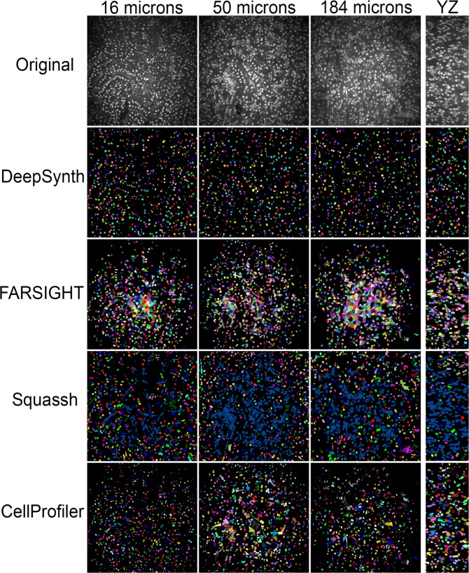
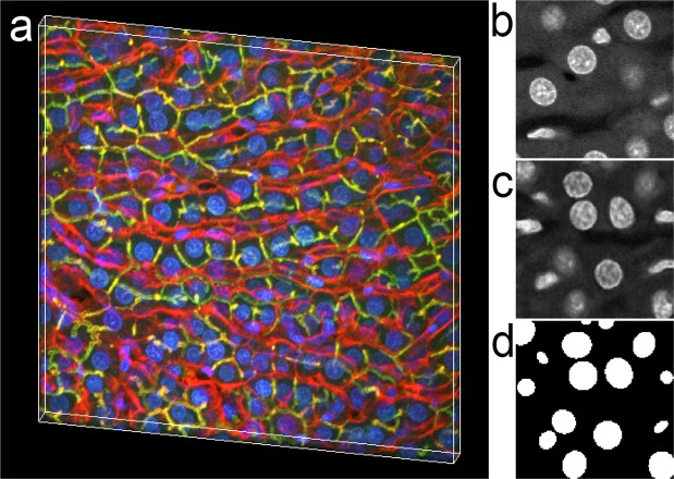
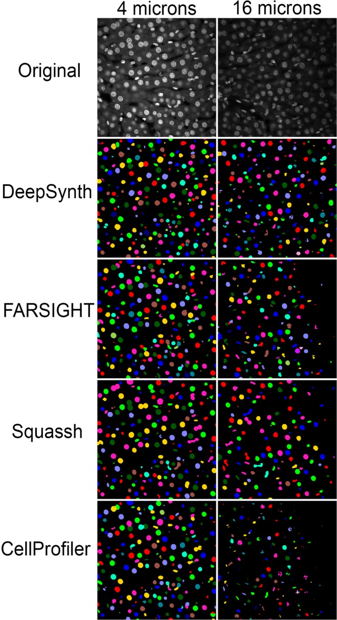
The scale of biological microscopy has increased dramatically over the past ten years, with the development of new modalities supporting collection of high-resolution fluorescence image volumes spanning hundreds of microns if not millimeters. The size and complexity of these volumes is such that quantitative analysis requires automated methods of image processing to identify and characterize individual cells. For many workflows, this process starts with segmentation of nuclei that, due to their ubiquity, ease-of-labeling and relatively simple structure, make them appealing targets for automated detection of individual cells. However, in the context of large, three-dimensional image volumes, nuclei present many challenges to automated segmentation, such that conventional approaches are seldom effective and/or robust. Techniques based upon deep-learning have shown great promise, but enthusiasm for applying these techniques is tempered by the need to generate training data, an arduous task, particularly in three dimensions. Here we present results of a new technique of nuclear segmentation using neural networks trained on synthetic data. Comparisons with results obtained using commonly-used image processing packages demonstrate that DeepSynth provides the superior results associated with deep-learning techniques without the need for manual annotation.

摘要

在过去的十年中,生物显微镜的规模急剧扩大,新的模式支持收集高分辨率荧光图像体积,这些图像体积可以跨越数百微米甚至毫米。这些体积的大小和复杂性使得定量分析需要自动化的图像处理方法来识别和描述单个细胞。对于许多工作流程,这个过程始于细胞核的分割,由于细胞核的普遍性、易于标记和相对简单的结构,使得它们成为自动检测单个细胞的有吸引力的目标。然而,在大型三维图像体积的情况下,细胞核给自动化分割带来了许多挑战,以至于传统方法很少有效和/或鲁棒。基于深度学习的技术显示出了巨大的潜力,但由于需要生成训练数据,这是一项艰巨的任务,特别是在三维方面,应用这些技术的热情受到了抑制。在这里,我们展示了一种使用合成数据训练神经网络的新细胞核分割技术的结果。与使用常用图像处理软件包获得的结果进行比较表明,DeepSynth 提供了与深度学习技术相关的优越结果,而无需手动注释。

https://cdn.ncbi.nlm.nih.gov/pmc/blobs/3fe9/6892824/05b1ab6afaf6/41598_2019_54244_Fig7_HTML.jpg
https://cdn.ncbi.nlm.nih.gov/pmc/blobs/3fe9/6892824/b68d168ea676/41598_2019_54244_Fig1_HTML.jpg
https://cdn.ncbi.nlm.nih.gov/pmc/blobs/3fe9/6892824/f1d79c69a7af/41598_2019_54244_Fig2_HTML.jpg
https://cdn.ncbi.nlm.nih.gov/pmc/blobs/3fe9/6892824/073c20cb14a3/41598_2019_54244_Fig3_HTML.jpg
https://cdn.ncbi.nlm.nih.gov/pmc/blobs/3fe9/6892824/9464cc69b4fe/41598_2019_54244_Fig4_HTML.jpg
https://cdn.ncbi.nlm.nih.gov/pmc/blobs/3fe9/6892824/fe7a7d322a88/41598_2019_54244_Fig5_HTML.jpg
https://cdn.ncbi.nlm.nih.gov/pmc/blobs/3fe9/6892824/23c6e5812028/41598_2019_54244_Fig6_HTML.jpg
https://cdn.ncbi.nlm.nih.gov/pmc/blobs/3fe9/6892824/05b1ab6afaf6/41598_2019_54244_Fig7_HTML.jpg

相似文献

1
DeepSynth: Three-dimensional nuclear segmentation of biological images using neural networks trained with synthetic data.DeepSynth:使用合成数据训练的神经网络进行生物图像的三维核分割。
Sci Rep. 2019 Dec 4;9(1):18295. doi: 10.1038/s41598-019-54244-5.
2
NISNet3D: three-dimensional nuclear synthesis and instance segmentation for fluorescence microscopy images.NISNet3D:用于荧光显微镜图像的三维核合成和实例分割。
Sci Rep. 2023 Jun 12;13(1):9533. doi: 10.1038/s41598-023-36243-9.
3
Application of convolutional neural networks towards nuclei segmentation in localization-based super-resolution fluorescence microscopy images.基于定位的超分辨率荧光显微镜图像中核分割的卷积神经网络应用。
BMC Bioinformatics. 2021 Jun 15;22(1):325. doi: 10.1186/s12859-021-04245-x.
4
Automatic image annotation for fluorescent cell nuclei segmentation.用于荧光细胞核分割的自动图像标注
PLoS One. 2021 Apr 16;16(4):e0250093. doi: 10.1371/journal.pone.0250093. eCollection 2021.
5
A Deep Learning Pipeline for Nucleus Segmentation.一种用于细胞核分割的深度学习管道。
Cytometry A. 2020 Dec;97(12):1248-1264. doi: 10.1002/cyto.a.24257. Epub 2020 Nov 19.
6
Bi-channel image registration and deep-learning segmentation (BIRDS) for efficient, versatile 3D mapping of mouse brain.双通道图像配准和深度学习分割(BIRDS)用于高效、通用的小鼠大脑 3D 映射。
Elife. 2021 Jan 18;10:e63455. doi: 10.7554/eLife.63455.
7
Robust and automated three-dimensional segmentation of densely packed cell nuclei in different biological specimens with Lines-of-Sight decomposition.利用视线分解对不同生物样本中紧密堆积的细胞核进行稳健且自动化的三维分割。
BMC Bioinformatics. 2015 Jun 8;16:187. doi: 10.1186/s12859-015-0617-x.
8
Evaluation of Deep Learning Architectures for Complex Immunofluorescence Nuclear Image Segmentation.深度学习架构在复杂免疫荧光核图像分割中的评估。
IEEE Trans Med Imaging. 2021 Jul;40(7):1934-1949. doi: 10.1109/TMI.2021.3069558. Epub 2021 Jun 30.
9
Automatic three-dimensional segmentation of mouse embryonic stem cell nuclei by utilising multiple channels of confocal fluorescence images.利用共聚焦荧光图像的多个通道自动对小鼠胚胎干细胞核进行三维分割。
J Microsc. 2021 Jan;281(1):57-75. doi: 10.1111/jmi.12949. Epub 2020 Aug 8.
10
Cellpose: a generalist algorithm for cellular segmentation.Cellpose:一种通用的细胞分割算法。
Nat Methods. 2021 Jan;18(1):100-106. doi: 10.1038/s41592-020-01018-x. Epub 2020 Dec 14.

引用本文的文献

1
AnyStar: Domain randomized universal star-convex 3D instance segmentation.AnyStar:域随机化通用星凸3D实例分割
IEEE Winter Conf Appl Comput Vis. 2024 Jan;2024:7578-7588. doi: 10.1109/wacv57701.2024.00742. Epub 2024 Apr 9.
2
Distributed and networked analysis of volumetric image data for remote collaboration of microscopy image analysis.用于显微镜图像分析远程协作的体积图像数据的分布式和网络化分析。
J Med Imaging (Bellingham). 2025 Mar;12(2):024001. doi: 10.1117/1.JMI.12.2.024001. Epub 2025 Mar 11.
3
Improving 3D deep learning segmentation with biophysically motivated cell synthesis.

本文引用的文献

1
Evaluation of Deep Learning Strategies for Nucleus Segmentation in Fluorescence Images.深度学习策略在荧光图像中细胞核分割的评估。
Cytometry A. 2019 Sep;95(9):952-965. doi: 10.1002/cyto.a.23863. Epub 2019 Jul 16.
2
Deep Learning in Image Cytometry: A Review.深度学习在图像细胞检测中的应用综述。
Cytometry A. 2019 Apr;95(4):366-380. doi: 10.1002/cyto.a.23701. Epub 2018 Dec 19.
3
U-Net: deep learning for cell counting, detection, and morphometry.U-Net:用于细胞计数、检测和形态测量学的深度学习。
通过具有生物物理动机的细胞合成改进3D深度学习分割
Commun Biol. 2025 Jan 11;8(1):43. doi: 10.1038/s42003-025-07469-2.
4
A spheroid whole mount drug testing pipeline with machine-learning based image analysis identifies cell-type specific differences in drug efficacy on a single-cell level.一种基于机器学习图像分析的球体整装药物测试流程可在单细胞水平上识别药物疗效的细胞类型特异性差异。
BMC Cancer. 2024 Dec 18;24(1):1542. doi: 10.1186/s12885-024-13329-9.
5
Nuclei Detection and Segmentation of Histopathological Images Using a Feature Pyramidal Network Variant of a Mask R-CNN.使用Mask R-CNN的特征金字塔网络变体进行组织病理学图像的细胞核检测与分割
Bioengineering (Basel). 2024 Oct 1;11(10):994. doi: 10.3390/bioengineering11100994.
6
This Microtubule Does Not Exist: Super-Resolution Microscopy Image Generation by a Diffusion Model.此微管不存在:基于扩散模型的超分辨率显微镜图像生成
Small Methods. 2025 Mar;9(3):e2400672. doi: 10.1002/smtd.202400672. Epub 2024 Oct 14.
7
A multiparametric analysis including single-cell and subcellular feature assessment reveals differential behavior of spheroid cultures on distinct ultra-low attachment plate types.一项包括单细胞和亚细胞特征评估的多参数分析揭示了在不同的超低附着板类型上球体培养物的差异行为。
Front Bioeng Biotechnol. 2024 Aug 2;12:1422235. doi: 10.3389/fbioe.2024.1422235. eCollection 2024.
8
Universal consensus 3D segmentation of cells from 2D segmented stacks.从二维分割堆栈中对细胞进行通用一致的三维分割。
bioRxiv. 2025 Mar 20:2024.05.03.592249. doi: 10.1101/2024.05.03.592249.
9
3DCellComposer - A Versatile Pipeline Utilizing 2D Cell Segmentation Methods for 3D Cell Segmentation.3D细胞合成器——一种利用二维细胞分割方法进行三维细胞分割的通用流程。
bioRxiv. 2024 Oct 27:2024.03.08.584082. doi: 10.1101/2024.03.08.584082.
10
Schooling of light reflecting fish.光反射鱼的教育。
PLoS One. 2023 Jul 21;18(7):e0289026. doi: 10.1371/journal.pone.0289026. eCollection 2023.
Nat Methods. 2019 Jan;16(1):67-70. doi: 10.1038/s41592-018-0261-2. Epub 2018 Dec 17.
4
A high-throughput imaging and nuclear segmentation analysis protocol for cleared 3D culture models.高通量成像和核分割分析方案用于已清除的 3D 培养模型。
Sci Rep. 2018 Jul 24;8(1):11135. doi: 10.1038/s41598-018-29169-0.
5
Deep Learning in Microscopy Image Analysis: A Survey.深度学习在显微镜图像分析中的应用:综述。
IEEE Trans Neural Netw Learn Syst. 2018 Oct;29(10):4550-4568. doi: 10.1109/TNNLS.2017.2766168. Epub 2017 Nov 22.
6
CellProfiler 3.0: Next-generation image processing for biology.CellProfiler 3.0:生物学的下一代图像处理。
PLoS Biol. 2018 Jul 3;16(7):e2005970. doi: 10.1371/journal.pbio.2005970. eCollection 2018 Jul.
7
Opportunities and obstacles for deep learning in biology and medicine.深度学习在生物学和医学中的机遇与挑战。
J R Soc Interface. 2018 Apr;15(141). doi: 10.1098/rsif.2017.0387.
8
Tamm-Horsfall Protein Regulates Mononuclear Phagocytes in the Kidney.Tamm-Horsfall 蛋白在肾脏中调节单核吞噬细胞。
J Am Soc Nephrol. 2018 Mar;29(3):841-856. doi: 10.1681/ASN.2017040409. Epub 2017 Nov 27.
9
Multiplex, quantitative cellular analysis in large tissue volumes with clearing-enhanced 3D microscopy (C3D).利用增强清除的 3D 显微镜(C3D)进行多重、定量细胞分析在大组织体积中。
Proc Natl Acad Sci U S A. 2017 Aug 29;114(35):E7321-E7330. doi: 10.1073/pnas.1708981114. Epub 2017 Aug 14.
10
Automated Training of Deep Convolutional Neural Networks for Cell Segmentation.自动化的深度学习卷积神经网络用于细胞分割。
Sci Rep. 2017 Aug 10;7(1):7860. doi: 10.1038/s41598-017-07599-6.