Department of Radiology, Seoul National University Hospital, Seoul, Korea.
Department of Radiology, Seoul National University College of Medicine, Seoul, Korea.
Korean J Radiol. 2019 Jul;20(7):1019-1041. doi: 10.3348/kjr.2018.0636.
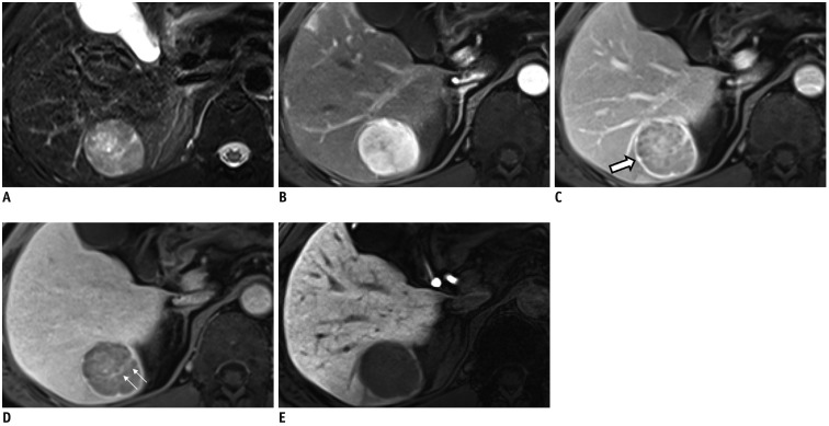
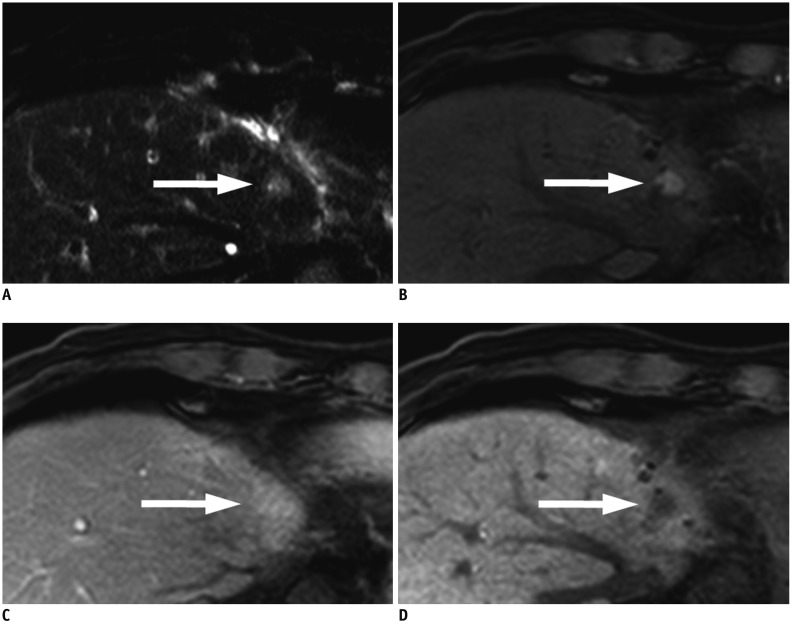
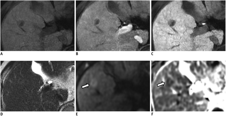
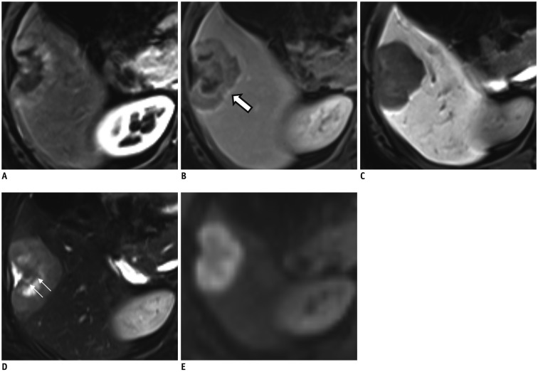
Hepatocellular carcinoma (HCC) can be diagnosed noninvasively with contrast-enhanced dynamic computed tomography, magnetic resonance imaging, or ultrasonography on the basis of its hallmark imaging features of arterial phase hyperenhancement and washout on portal or delayed phase images. However, approximately 40% of HCCs show atypical imaging features, posing a significant diagnostic challenge for radiologists. Another challenge for radiologists in clinical practice is the presentation of many HCC mimickers such as intrahepatic cholangiocarcinoma, combined HCC-cholangiocarcinoma, arterioportal shunt, and hemangioma in the cirrhotic liver. The differentiation of HCCs from these mimickers on preoperative imaging studies is of critical importance. Hence, we will review the typical and atypical imaging features of HCCs and the imaging features of its common mimickers. In addition, we will discuss how to solve these challenges in practice.
肝细胞癌 (HCC) 可以通过增强动态计算机断层扫描、磁共振成像或超声检查,根据其标志性的影像学特征,即动脉期增强和门静脉或延迟期图像的洗脱来进行非侵入性诊断。然而,约 40%的 HCC 表现出不典型的影像学特征,这对放射科医生来说是一个重大的诊断挑战。放射科医生在临床实践中面临的另一个挑战是,许多 HCC 的类似物,如肝内胆管细胞癌、合并 HCC-胆管细胞癌、动静脉分流和肝硬化中的血管瘤,在术前影像学研究中表现出类似 HCC 的特征。因此,术前影像学研究中区分 HCC 与这些类似物至关重要。因此,我们将回顾 HCC 的典型和不典型影像学特征及其常见类似物的影像学特征。此外,我们将讨论如何在实践中解决这些挑战。