• 文献检索
  • 文档翻译
  • 深度研究
  • 学术资讯
  • Suppr Zotero 插件Zotero 插件
  • 邀请有礼
  • 套餐&价格
  • 历史记录
应用&插件
Suppr Zotero 插件Zotero 插件浏览器插件Mac 客户端Windows 客户端微信小程序
定价
高级版会员购买积分包购买API积分包
服务
文献检索文档翻译深度研究API 文档MCP 服务
关于我们
关于 Suppr公司介绍联系我们用户协议隐私条款
关注我们

Suppr 超能文献

核心技术专利:CN118964589B侵权必究
粤ICP备2023148730 号-1Suppr @ 2026

文献检索

告别复杂PubMed语法,用中文像聊天一样搜索,搜遍4000万医学文献。AI智能推荐,让科研检索更轻松。

立即免费搜索

文件翻译

保留排版,准确专业,支持PDF/Word/PPT等文件格式,支持 12+语言互译。

免费翻译文档

深度研究

AI帮你快速写综述,25分钟生成高质量综述,智能提取关键信息,辅助科研写作。

立即免费体验

青光眼患者视白质通路的多对比度磁共振成像。

Multi-Contrast Magnetic Resonance Imaging of Visual White Matter Pathways in Patients With Glaucoma.

机构信息

Department of Ophthalmology, The Jikei University School of Medicine, Tokyo, Japan.

Center for Information and Neural Networks (CiNet), Advanced ICT Research Institute, National Institute of Information and Communications Technology, Suita, Japan.

出版信息

Invest Ophthalmol Vis Sci. 2022 Feb 1;63(2):29. doi: 10.1167/iovs.63.2.29.

DOI:10.1167/iovs.63.2.29
PMID:35201263
原文链接:https://pmc.ncbi.nlm.nih.gov/articles/PMC8883150/
Abstract

PURPOSE

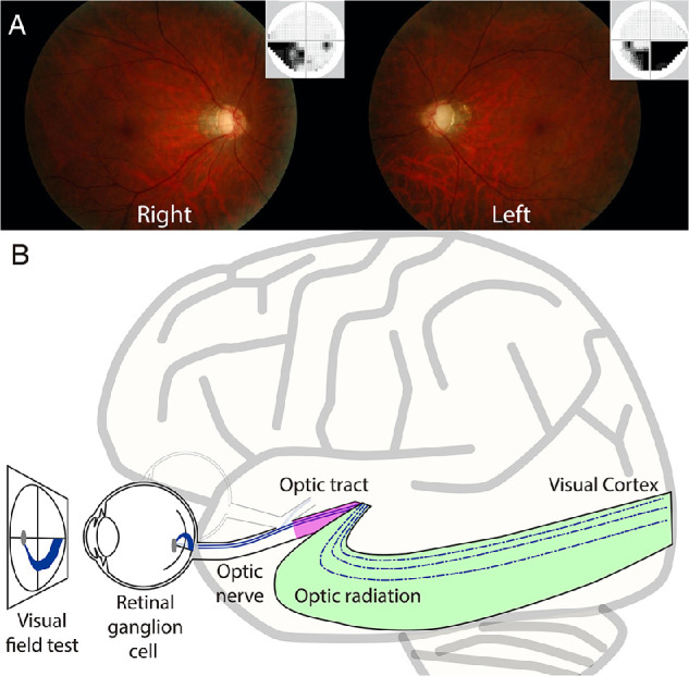
Glaucoma is a disorder that involves visual field loss caused by retinal ganglion cell damage. Previous diffusion magnetic resonance imaging (dMRI) studies have demonstrated that retinal ganglion cell damage affects tissues in the optic tract (OT) and optic radiation (OR). However, because previous studies have used a simple diffusion tensor model to analyze dMRI data, the microstructural interpretation of white matter tissue changes remains uncertain. In this study, we used a multi-contrast MRI approach to further clarify the type of microstructural damage that occurs in patients with glaucoma.

METHODS

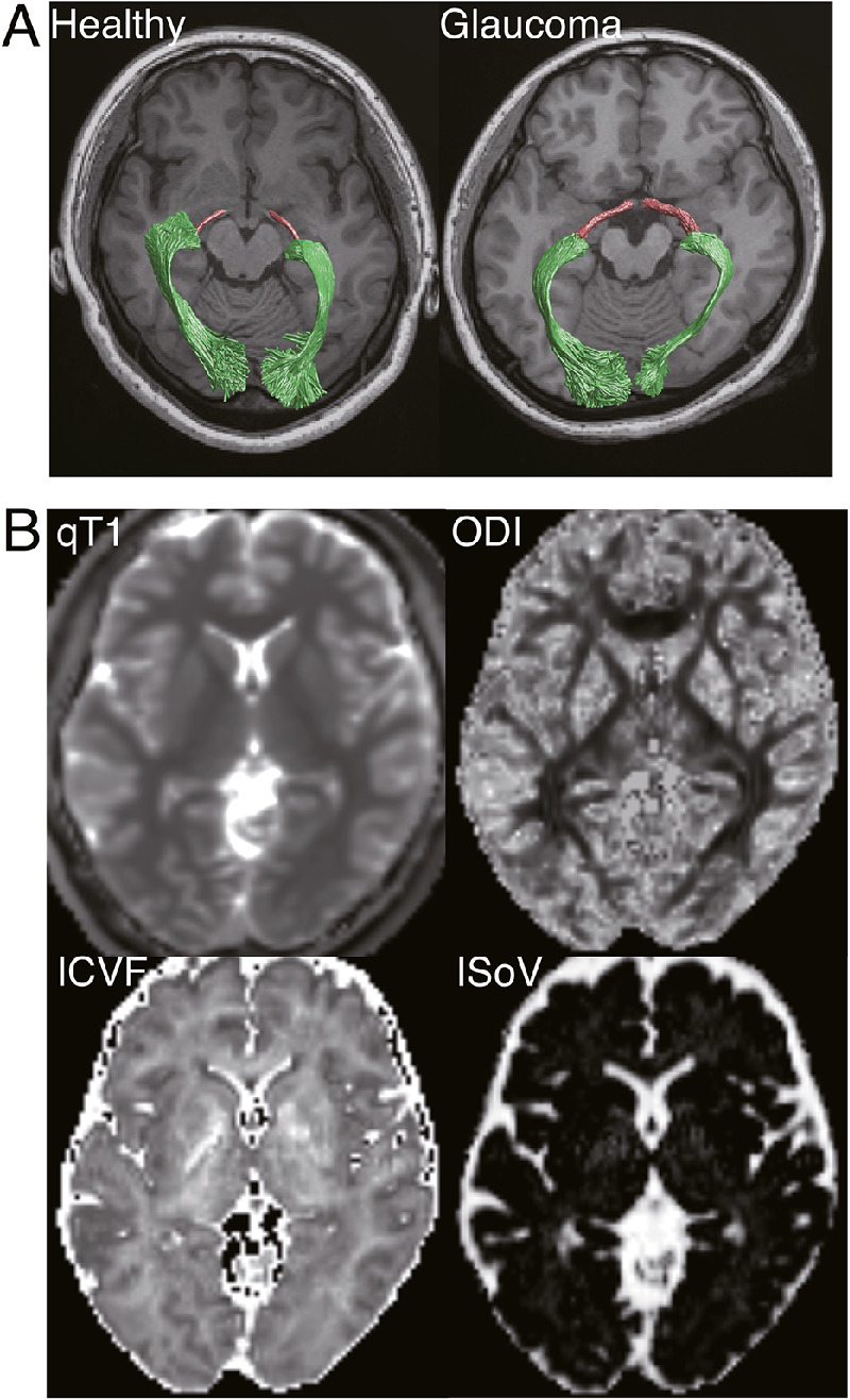
We collected dMRI data from 17 patients with glaucoma and 30 controls using 3-tesla (3T) MRI. Using the dMRI data, we estimated three types of tissue property metrics: intracellular volume fraction (ICVF), orientation dispersion index (ODI), and isotropic volume fraction (IsoV). Quantitative T1 (qT1) data, which may be relatively specific to myelin, were collected from all subjects.

RESULTS

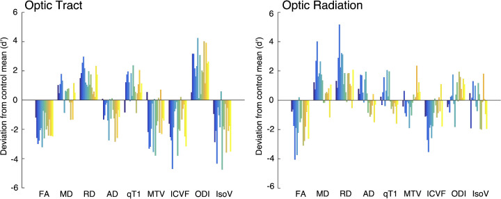
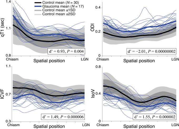
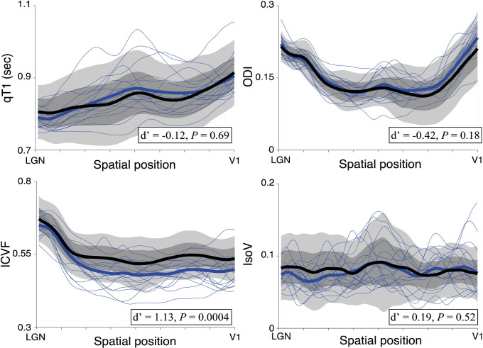
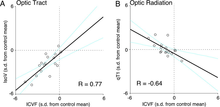
In the OT, all four metrics showed significant differences between the glaucoma and control groups. In the OR, only the ICVF showed significant between-group differences. ICVF was significantly correlated with qT1 in the OR of the glaucoma group, although qT1 did not show any abnormality at the group level.

CONCLUSIONS

Our results suggest that, at the group level, tissue changes in OR caused by glaucoma might be explained by axonal damage, which is reflected in the intracellular diffusion signals, rather than myelin damage. The significant correlation between ICVF and qT1 suggests that myelin damage might also occur in a smaller number of severe cases.

摘要

目的

青光眼是一种涉及视网膜神经节细胞损伤导致视野丧失的疾病。先前的扩散磁共振成像(dMRI)研究表明,视网膜神经节细胞损伤会影响视束(OT)和视辐射(OR)中的组织。然而,由于先前的研究使用简单的扩散张量模型来分析 dMRI 数据,因此对白质组织变化的微观结构解释仍不确定。在这项研究中,我们使用多对比度 MRI 方法进一步阐明青光眼患者中发生的微观结构损伤的类型。

方法

我们使用 3 特斯拉(3T)MRI 从 17 名青光眼患者和 30 名对照者中收集了 dMRI 数据。使用 dMRI 数据,我们估计了三种组织属性指标:细胞内体积分数(ICVF)、方向分散指数(ODI)和各向同性体积分数(IsoV)。从所有受试者中收集了定量 T1(qT1)数据,该数据可能与髓鞘相对特异。

结果

在 OT 中,四项指标在青光眼组和对照组之间均显示出显著差异。在 OR 中,只有 ICVF 显示出组间差异。尽管 qT1 在组水平上没有显示任何异常,但 ICVF 与青光眼组 OR 中的 qT1 呈显著相关。

结论

我们的结果表明,在组水平上,青光眼引起的 OR 中的组织变化可能是由轴突损伤引起的,这反映在细胞内扩散信号中,而不是髓鞘损伤。ICVF 与 qT1 之间的显著相关性表明,髓鞘损伤也可能在少数严重病例中发生。

https://cdn.ncbi.nlm.nih.gov/pmc/blobs/b054/8883150/f0056abc3a22/iovs-63-2-29-f006.jpg
https://cdn.ncbi.nlm.nih.gov/pmc/blobs/b054/8883150/cc5f4c3b15e5/iovs-63-2-29-f001.jpg
https://cdn.ncbi.nlm.nih.gov/pmc/blobs/b054/8883150/adeae034349f/iovs-63-2-29-f002.jpg
https://cdn.ncbi.nlm.nih.gov/pmc/blobs/b054/8883150/060ba722fcba/iovs-63-2-29-f003.jpg
https://cdn.ncbi.nlm.nih.gov/pmc/blobs/b054/8883150/30fe46dfa054/iovs-63-2-29-f004.jpg
https://cdn.ncbi.nlm.nih.gov/pmc/blobs/b054/8883150/6cf3e7dd1586/iovs-63-2-29-f005.jpg
https://cdn.ncbi.nlm.nih.gov/pmc/blobs/b054/8883150/f0056abc3a22/iovs-63-2-29-f006.jpg

相似文献

1
Multi-Contrast Magnetic Resonance Imaging of Visual White Matter Pathways in Patients With Glaucoma.青光眼患者视白质通路的多对比度磁共振成像。
Invest Ophthalmol Vis Sci. 2022 Feb 1;63(2):29. doi: 10.1167/iovs.63.2.29.
2
Diffusivity and quantitative T1 profile of human visual white matter tracts after retinal ganglion cell damage.人视网膜神经节细胞损伤后视觉白质束的扩散率和定量 T1 谱。
Neuroimage Clin. 2019;23:101826. doi: 10.1016/j.nicl.2019.101826. Epub 2019 Apr 16.
3
Microstructural Visual Pathway White Matter Alterations in Primary Open-Angle Glaucoma: A Neurite Orientation Dispersion and Density Imaging Study.原发性开角型青光眼微观结构视觉通路白质改变:神经丝取向分散和密度成像研究。
AJNR Am J Neuroradiol. 2022 May;43(5):756-763. doi: 10.3174/ajnr.A7495. Epub 2022 Apr 21.
4
Axonal damage in the optic radiation assessed by white matter tract integrity metrics is associated with retinal thinning in multiple sclerosis.轴突损伤在评估视辐射中的白质束完整性指标与多发性硬化症中的视网膜变薄有关。
Neuroimage Clin. 2020;27:102293. doi: 10.1016/j.nicl.2020.102293. Epub 2020 May 26.
5
Contributions of Brain Microstructures and Metabolism to Visual Field Loss Patterns in Glaucoma Using Archetypal and Information Gain Analyses.利用原型和信息增益分析探讨脑微结构和代谢对青光眼视野丧失模式的影响。
Invest Ophthalmol Vis Sci. 2024 Jul 1;65(8):15. doi: 10.1167/iovs.65.8.15.
6
Fixel-Based Analysis of Visual Pathway White Matter in Primary Open-Angle Glaucoma.基于体素的原发性开角型青光眼视路白质分析。
Invest Ophthalmol Vis Sci. 2019 Sep 3;60(12):3803-3812. doi: 10.1167/iovs.19-27447.
7
Diffusion tensor magnetic resonance imaging reveals visual pathway damage that correlates with clinical severity in glaucoma.弥散张量磁共振成像显示视路损伤与青光眼临床严重程度相关。
Clin Exp Ophthalmol. 2013 Jan-Feb;41(1):43-9. doi: 10.1111/j.1442-9071.2012.02832.x. Epub 2012 Aug 21.
8
White and Gray Matter Abnormality in Burning Mouth Syndrome Evaluated with Diffusion Tensor Imaging and Neurite Orientation Dispersion and Density Imaging.弥散张量成像和神经丝取向分散和密度成像评估灼口综合征的白质和灰质异常。
Magn Reson Med Sci. 2024 Apr 1;23(2):204-213. doi: 10.2463/mrms.mp.2022-0099. Epub 2023 Mar 29.
9
Assessing Visual Pathway White Matter Degeneration in Primary Open-Angle Glaucoma Using Multiple MRI Morphology and Diffusion Metrics.使用多种MRI形态学和扩散指标评估原发性开角型青光眼的视觉通路白质退变
J Magn Reson Imaging. 2025 Apr;61(4):1699-1711. doi: 10.1002/jmri.29616. Epub 2024 Sep 23.
10
Optic Radiations Microstructural Changes in Glaucoma and Association With Severity: A Study Using 3Tesla-Magnetic Resonance Diffusion Tensor Imaging.青光眼视辐射的微观结构变化及其与严重程度的关联:一项使用3特斯拉磁共振扩散张量成像的研究
Invest Ophthalmol Vis Sci. 2016 Dec 1;57(15):6539-6547. doi: 10.1167/iovs.16-19838.

引用本文的文献

1
Evaluating the impact of denoising diffusion MRI data on tractometry metrics of optic tract abnormalities in glaucoma.评估去噪扩散磁共振成像数据对青光眼视束异常的纤维束测量指标的影响。
Sci Rep. 2025 Jul 16;15(1):25812. doi: 10.1038/s41598-025-10947-6.
2
Associations Between Myopia and Brain Volumes: An Observational and Genetic Analysis.近视与脑容量之间的关联:一项观察性与遗传学分析。
Invest Ophthalmol Vis Sci. 2025 Jun 2;66(6):57. doi: 10.1167/iovs.66.6.57.
3
Application of advanced diffusion MRI based tractometry of the visual pathway in glaucoma: a systematic review.

本文引用的文献

1
Dissecting whole-brain conduction delays through MRI microstructural measures.通过 MRI 微观结构测量解析全脑传导延迟。
Brain Struct Funct. 2021 Nov;226(8):2651-2663. doi: 10.1007/s00429-021-02358-w. Epub 2021 Aug 14.
2
Age dependency and lateralization in the three branches of the human superior longitudinal fasciculus.人类上纵束三支的年龄依赖性和侧化。
Cortex. 2021 Jun;139:116-133. doi: 10.1016/j.cortex.2021.02.027. Epub 2021 Mar 15.
3
White matter alterations in glaucoma and monocular blindness differ outside the visual system.
基于先进扩散磁共振成像的视路纤维束成像技术在青光眼诊断中的应用:一项系统评价
Front Neurosci. 2025 May 21;19:1577991. doi: 10.3389/fnins.2025.1577991. eCollection 2025.
4
Glaucoma and brain functional networks: a bidirectional Mendelian randomisation study.青光眼与脑功能网络:一项双向孟德尔随机化研究
BMJ Open Ophthalmol. 2025 Apr 12;10(1):e001902. doi: 10.1136/bmjophth-2024-001902.
5
Association Between Glaucoma and Brain Structural Connectivity Based on Diffusion Tensor Tractography: A Bidirectional Mendelian Randomization Study.基于扩散张量纤维束成像的青光眼与脑结构连通性之间的关联:一项双向孟德尔随机化研究
Brain Sci. 2024 Oct 17;14(10):1030. doi: 10.3390/brainsci14101030.
6
Causal Relationship between Aging and Anorexia Nervosa: A White-Matter-Microstructure-Mediated Mendelian Randomization Analysis.衰老与神经性厌食症之间的因果关系:一项基于白质微结构介导的孟德尔随机化分析
Biomedicines. 2024 Aug 16;12(8):1874. doi: 10.3390/biomedicines12081874.
7
Tractometry of Human Visual White Matter Pathways in Health and Disease.人类视觉白质通路的束路测量:在健康与疾病中的应用
Magn Reson Med Sci. 2024 Jul 1;23(3):316-340. doi: 10.2463/mrms.rev.2024-0007. Epub 2024 Jun 12.
青光眼和单眼失明患者视觉系统之外的白质改变存在差异。
Sci Rep. 2021 Mar 25;11(1):6866. doi: 10.1038/s41598-021-85602-x.
4
An interactive meta-analysis of MRI biomarkers of myelin.MRI 生物标志物的交互元分析:髓鞘
Elife. 2020 Oct 21;9:e61523. doi: 10.7554/eLife.61523.
5
Diffusion Kurtosis Imaging Reveals Optic Tract Damage That Correlates with Clinical Severity in Glaucoma.扩散峰度成像揭示青光眼视束损伤与临床严重程度相关。
Annu Int Conf IEEE Eng Med Biol Soc. 2020 Jul;2020:1746-1749. doi: 10.1109/EMBC44109.2020.9176192.
6
NODDI in clinical research.临床研究中的神经突方向离散与密度成像(NODDI)
J Neurosci Methods. 2020 Dec 1;346:108908. doi: 10.1016/j.jneumeth.2020.108908. Epub 2020 Aug 16.
7
Imaging G-Ratio in Multiple Sclerosis Using High-Gradient Diffusion MRI and Macromolecular Tissue Volume.使用高梯度弥散 MRI 和大分子组织容积成像技术评估多发性硬化症的 G 比值
AJNR Am J Neuroradiol. 2019 Nov;40(11):1871-1877. doi: 10.3174/ajnr.A6283. Epub 2019 Nov 6.
8
Normal tension glaucoma-like degeneration of the visual system in aged marmosets.老年狨猴视觉系统的正常眼压样变性。
Sci Rep. 2019 Oct 16;9(1):14852. doi: 10.1038/s41598-019-51281-y.
9
Quantification of apparent axon density and orientation dispersion in the white matter of youth born with congenital heart disease.量化先天性心脏病患儿白质中表观轴突密度和方向分散度。
Neuroimage. 2020 Jan 15;205:116255. doi: 10.1016/j.neuroimage.2019.116255. Epub 2019 Oct 9.
10
Fixel-Based Analysis of Visual Pathway White Matter in Primary Open-Angle Glaucoma.基于体素的原发性开角型青光眼视路白质分析。
Invest Ophthalmol Vis Sci. 2019 Sep 3;60(12):3803-3812. doi: 10.1167/iovs.19-27447.