• 文献检索
  • 文档翻译
  • 深度研究
  • 学术资讯
  • Suppr Zotero 插件Zotero 插件
  • 邀请有礼
  • 套餐&价格
  • 历史记录
应用&插件
Suppr Zotero 插件Zotero 插件浏览器插件Mac 客户端Windows 客户端微信小程序
定价
高级版会员购买积分包购买API积分包
服务
文献检索文档翻译深度研究API 文档MCP 服务
关于我们
关于 Suppr公司介绍联系我们用户协议隐私条款
关注我们

Suppr 超能文献

核心技术专利:CN118964589B侵权必究
粤ICP备2023148730 号-1Suppr @ 2026

文献检索

告别复杂PubMed语法,用中文像聊天一样搜索,搜遍4000万医学文献。AI智能推荐,让科研检索更轻松。

立即免费搜索

文件翻译

保留排版,准确专业,支持PDF/Word/PPT等文件格式,支持 12+语言互译。

免费翻译文档

深度研究

AI帮你快速写综述,25分钟生成高质量综述,智能提取关键信息,辅助科研写作。

立即免费体验

下腔静脉边缘追踪超声心动图:一种在多种临床场景中具有应用前景的工具。

Inferior Vena Cava Edge Tracking Echocardiography: A Promising Tool with Applications in Multiple Clinical Settings.

作者信息

Albani Stefano, Mesin Luca, Roatta Silvestro, De Luca Antonio, Giannoni Alberto, Stolfo Davide, Biava Lorenza, Bonino Caterina, Contu Laura, Pelloni Elisa, Attena Emilio, Russo Vincenzo, Antonini-Canterin Francesco, Pugliese Nicola Riccardo, Gallone Guglielmo, De Ferrari Gaetano Maria, Sinagra Gianfranco, Scacciatella Paolo

机构信息

Division of Cardiology, Umberto Parini Regional Hospital, 11100 Aosta, Italy.

Cardio-Thoraco-Vascular Department, Division of Cardiology and Postgraduate School in Cardiovascular Sciences, University of Trieste, 34127 Trieste, Italy.

出版信息

Diagnostics (Basel). 2022 Feb 7;12(2):427. doi: 10.3390/diagnostics12020427.

DOI:10.3390/diagnostics12020427
PMID:35204518
原文链接:https://pmc.ncbi.nlm.nih.gov/articles/PMC8871248/
Abstract

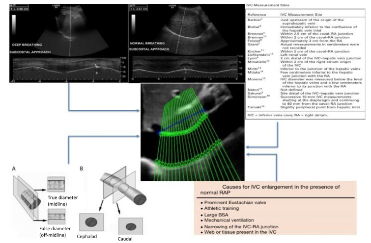
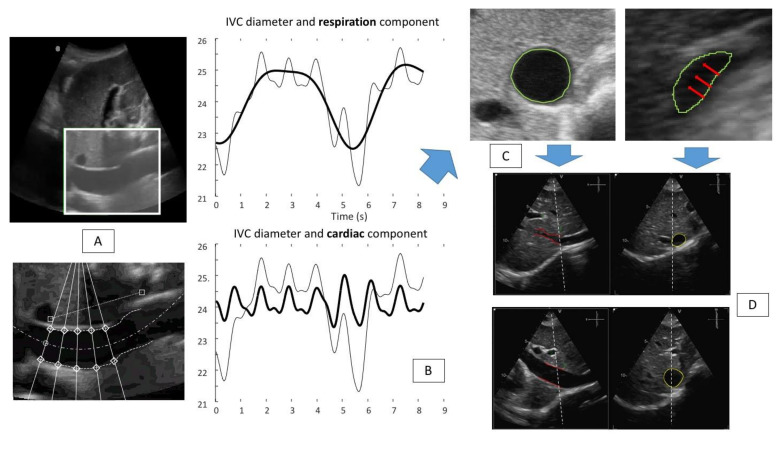
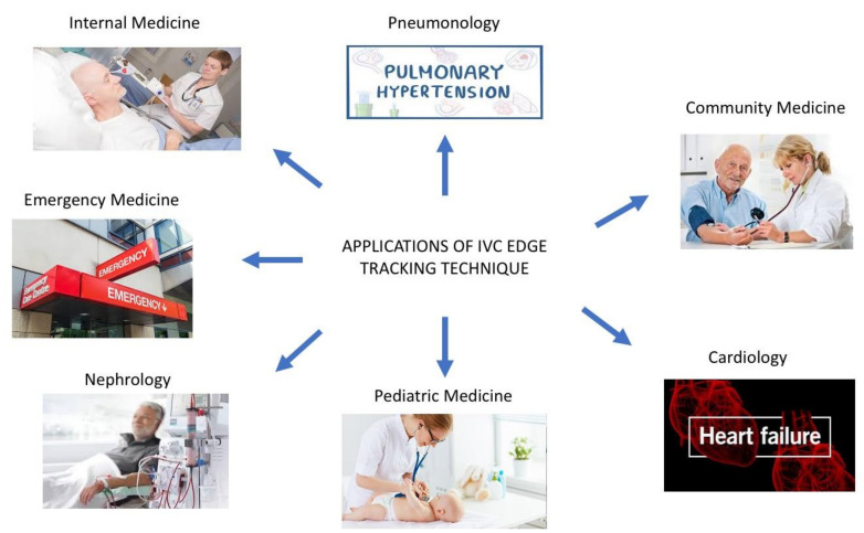
Ultrasound (US)-based measurements of the inferior vena cava (IVC) diameter are widely used to estimate right atrial pressure (RAP) in a variety of clinical settings. However, the correlation with invasively measured RAP along with the reproducibility of US-based IVC measurements is modest at best. In the present manuscript, we discuss the limitations of the current technique to estimate RAP through IVC US assessment and present a new promising tool developed by our research group, the automated IVC edge-to-edge tracking system, which has the potential to improve RAP assessment by transforming the current categorical classification (low, normal, high RAP) in a continuous and precise RAP estimation technique. Finally, we critically evaluate all the clinical settings in which this new tool could improve current practice.

摘要

基于超声(US)测量下腔静脉(IVC)直径在各种临床环境中被广泛用于估计右心房压力(RAP)。然而,与通过侵入性测量获得的RAP之间的相关性以及基于US的IVC测量的可重复性充其量只是中等程度。在本手稿中,我们讨论了通过IVC US评估来估计RAP的当前技术的局限性,并介绍了我们研究小组开发的一种新的有前景的工具——自动IVC边缘到边缘跟踪系统,该系统有可能通过将当前的分类(低、正常、高RAP)转变为一种连续且精确的RAP估计技术来改善RAP评估。最后,我们严格评估了这种新工具能够改进当前实践的所有临床环境。

https://cdn.ncbi.nlm.nih.gov/pmc/blobs/632a/8871248/413c404639e3/diagnostics-12-00427-g005.jpg
https://cdn.ncbi.nlm.nih.gov/pmc/blobs/632a/8871248/7c291a71f873/diagnostics-12-00427-g001.jpg
https://cdn.ncbi.nlm.nih.gov/pmc/blobs/632a/8871248/c30944b80565/diagnostics-12-00427-g002.jpg
https://cdn.ncbi.nlm.nih.gov/pmc/blobs/632a/8871248/69712d877a4d/diagnostics-12-00427-g003.jpg
https://cdn.ncbi.nlm.nih.gov/pmc/blobs/632a/8871248/be52e92e3695/diagnostics-12-00427-g004.jpg
https://cdn.ncbi.nlm.nih.gov/pmc/blobs/632a/8871248/413c404639e3/diagnostics-12-00427-g005.jpg

相似文献

1
Inferior Vena Cava Edge Tracking Echocardiography: A Promising Tool with Applications in Multiple Clinical Settings.下腔静脉边缘追踪超声心动图:一种在多种临床场景中具有应用前景的工具。
Diagnostics (Basel). 2022 Feb 7;12(2):427. doi: 10.3390/diagnostics12020427.
2
Accuracy of right atrial pressure estimation using a multi-parameter approach derived from inferior vena cava semi-automated edge-tracking echocardiography: a pilot study in patients with cardiovascular disorders.应用下腔静脉半自动边缘追踪超声心动图多参数法评估右心房压力的准确性:心血管疾病患者的初步研究。
Int J Cardiovasc Imaging. 2020 Jul;36(7):1213-1225. doi: 10.1007/s10554-020-01814-8. Epub 2020 Mar 19.
3
Non-Invasive Estimation of Right Atrial Pressure Using a Semi-Automated Echocardiographic Tool for Inferior Vena Cava Edge-Tracking.使用半自动超声心动图工具进行下腔静脉边缘追踪对右心房压力的无创估计
J Clin Med. 2022 Jun 7;11(12):3257. doi: 10.3390/jcm11123257.
4
Evaluation of an artificial intelligence-based system for echocardiographic estimation of right atrial pressure.基于人工智能的超声心动图右心房压估测系统的评估。
Int J Cardiovasc Imaging. 2023 Dec;39(12):2437-2450. doi: 10.1007/s10554-023-02941-8. Epub 2023 Sep 8.
5
Coronary sinus collapsibility index and inferior vena cava collapsibility index can predict right atrial pressure in patients undergoing right heart catheterization.冠状窦塌陷指数和下腔静脉塌陷指数可预测行右心导管检查患者的右心房压。
Echocardiography. 2022 Mar;39(3):440-446. doi: 10.1111/echo.15314. Epub 2022 Feb 3.
6
Impact of Body Size on Inferior Vena Cava Parameters for Estimating Right Atrial Pressure: A Need for Standardization?体型对评估右心房压力的下腔静脉参数的影响:是否需要标准化?
J Am Soc Echocardiogr. 2015 Dec;28(12):1420-7. doi: 10.1016/j.echo.2015.07.008. Epub 2015 Aug 10.
7
Role of Inferior Vena Cava Dynamics for Estimating Right Atrial Pressure in Congenital Heart Disease.下腔静脉动力学在先天性心脏病右心房压力评估中的作用。
Circ Cardiovasc Imaging. 2022 Sep;15(9):e014308. doi: 10.1161/CIRCIMAGING.122.014308. Epub 2022 Sep 20.
8
Non-invasive Estimation of Right Atrial Pressure Using Inferior Vena Cava Echography.应用下腔静脉超声心动图无创估计右心房压力。
Ultrasound Med Biol. 2019 May;45(5):1331-1337. doi: 10.1016/j.ultrasmedbio.2018.12.013. Epub 2019 Feb 25.
9
Non-invasive estimation of right atrial pressure by combined Doppler echocardiographic measurements of the inferior vena cava in patients with congestive heart failure.通过联合多普勒超声心动图测量充血性心力衰竭患者下腔静脉来无创估计右心房压力。
Ital Heart J. 2000 Oct;1(10):684-90.
10
3D echocardiography to evaluate right atrial pressure in acutely decompensated heart failure correlation with invasive hemodynamics.三维超声心动图评价急性失代偿性心力衰竭患者右心房压与有创血流动力学的相关性。
JACC Cardiovasc Imaging. 2011 Sep;4(9):938-45. doi: 10.1016/j.jcmg.2011.05.006.

引用本文的文献

1
Prolapsing Intrigue: A Case of Superior Vena Cava Mass Visualized by Echocardiography From the Subcostal Window Unveiling an Anterior Mediastinal Type B2 Thymoma.脱垂之谜:一例通过肋下窗超声心动图显示的上腔静脉肿块,揭示了前纵隔B2型胸腺瘤
Cureus. 2024 Jan 16;16(1):e52352. doi: 10.7759/cureus.52352. eCollection 2024 Jan.
2
Refining accuracy of RV-PA coupling in patients undergoing transcatheter tricuspid valve treatment.优化经导管三尖瓣治疗患者的右心室-肺动脉偶联精准度。
Clin Res Cardiol. 2024 Jan;113(1):177-186. doi: 10.1007/s00392-023-02339-5. Epub 2023 Nov 27.
3
Processing Ultrasound Scans of the Inferior Vena Cava: Techniques and Applications.

本文引用的文献

1
Assessment of Phasic Changes of Vascular Size by Automated Edge Tracking-State of the Art and Clinical Perspectives.通过自动边缘跟踪评估血管大小的相位变化——最新技术与临床前景
Front Cardiovasc Med. 2022 Jan 21;8:775635. doi: 10.3389/fcvm.2021.775635. eCollection 2021.
2
The Cardiac Caval Index: Improving Noninvasive Assessment of Cardiac Preload.心腔指数:改善心脏前负荷的无创评估
J Ultrasound Med. 2022 Sep;41(9):2247-2258. doi: 10.1002/jum.15909. Epub 2021 Dec 8.
3
Multiparametric vs. Inferior Vena Cava-Based Estimation of Right Atrial Pressure.
下腔静脉超声扫描的处理:技术与应用
Bioengineering (Basel). 2023 Sep 12;10(9):1076. doi: 10.3390/bioengineering10091076.
4
Evaluation of an artificial intelligence-based system for echocardiographic estimation of right atrial pressure.基于人工智能的超声心动图右心房压估测系统的评估。
Int J Cardiovasc Imaging. 2023 Dec;39(12):2437-2450. doi: 10.1007/s10554-023-02941-8. Epub 2023 Sep 8.
5
Non-Invasive Estimation of Right Atrial Pressure Using a Semi-Automated Echocardiographic Tool for Inferior Vena Cava Edge-Tracking.使用半自动超声心动图工具进行下腔静脉边缘追踪对右心房压力的无创估计
J Clin Med. 2022 Jun 7;11(12):3257. doi: 10.3390/jcm11123257.
基于多参数与下腔静脉的右心房压力估计
Front Cardiovasc Med. 2021 Mar 8;8:632302. doi: 10.3389/fcvm.2021.632302. eCollection 2021.
4
A novel echocardiographic method for estimation of pulmonary artery wedge pressure and pulmonary vascular resistance.一种用于评估肺动脉楔压和肺血管阻力的新型超声心动图方法。
ESC Heart Fail. 2021 Apr;8(2):1216-1229. doi: 10.1002/ehf2.13183. Epub 2021 Feb 10.
5
Multi-directional Assessment of Respiratory and Cardiac Pulsatility of the Inferior Vena Cava From Ultrasound Imaging in Short Axis.超声短轴切面下下腔静脉呼吸及心脏搏动的多向性评估。
Ultrasound Med Biol. 2020 Dec;46(12):3475-3482. doi: 10.1016/j.ultrasmedbio.2020.08.027. Epub 2020 Sep 29.
6
Heart Failure With Reduced Ejection Fraction: A Review.射血分数降低型心力衰竭:综述。
JAMA. 2020 Aug 4;324(5):488-504. doi: 10.1001/jama.2020.10262.
7
Global Right Heart Assessment with Speckle-Tracking Imaging Improves the Risk Prediction of a Validated Scoring System in Pulmonary Arterial Hypertension.基于斑点追踪成像的全右心评估改善了肺动脉高压中经验证的评分系统的风险预测。
J Am Soc Echocardiogr. 2020 Nov;33(11):1334-1344.e2. doi: 10.1016/j.echo.2020.05.020. Epub 2020 Aug 1.
8
Feasibility of Using a Pocket-Sized Ultrasound Device to Measure the Inferior Vena Cava Diameter of Patients With Heart Failure in the Community Setting: A Pilot Study.在社区环境中使用口袋大小的超声设备测量心力衰竭患者下腔静脉直径的可行性:一项初步研究。
J Prim Care Community Health. 2020 Jan-Dec;11:2150132720931345. doi: 10.1177/2150132720931345.
9
Cardiac Variation of Internal Jugular Vein as a Marker of Volume Change in Hemorrhagic Shock.颈内静脉的心脏变异作为失血性休克容量变化的标志物
Shock. 2020 Dec;54(6):717-722. doi: 10.1097/SHK.0000000000001548.
10
Accuracy of right atrial pressure estimation using a multi-parameter approach derived from inferior vena cava semi-automated edge-tracking echocardiography: a pilot study in patients with cardiovascular disorders.应用下腔静脉半自动边缘追踪超声心动图多参数法评估右心房压力的准确性:心血管疾病患者的初步研究。
Int J Cardiovasc Imaging. 2020 Jul;36(7):1213-1225. doi: 10.1007/s10554-020-01814-8. Epub 2020 Mar 19.