Mokoala Kgomotso M G, Lawal Ismaheel O, Maserumule Letjie C, Hlongwa Khanyisile N, Ndlovu Honest, Reed Janet, Bida Meshack, Maes Alex, van de Wiele Christophe, Mahapane Johncy, Davis Cindy, Jeong Jae Min, Popoola Gbenga, Vorster Mariza, Sathekge Mike M
Department of Nuclear Medicine, University of Pretoria, Pretoria 0001, South Africa.
Nuclear Medicine Research Infrastructure (NuMeRI), Steve Biko Academic Hospital, Pretoria 0001, South Africa.
J Clin Med. 2022 Feb 12;11(4):962. doi: 10.3390/jcm11040962.
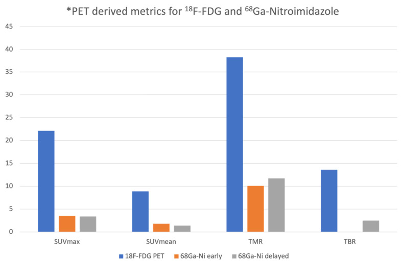
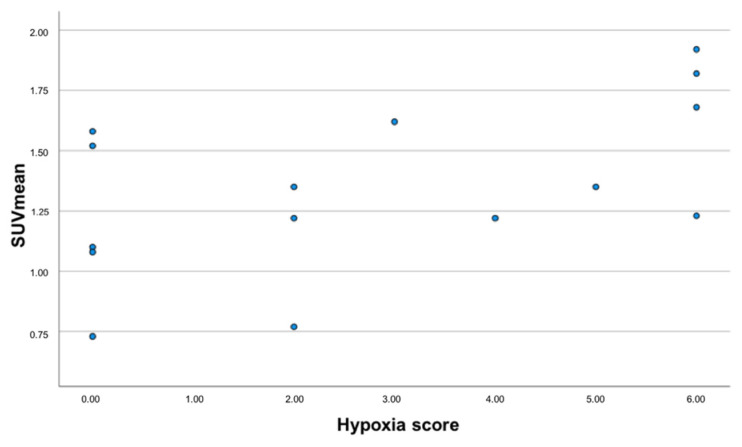
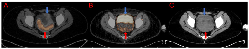
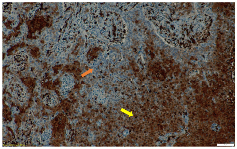
Hypoxia in cervical cancer has been associated with a poor prognosis. Over the years Ga labelled nitroimidazoles have been studied and have shown improved kinetics. We present our initial experience of hypoxia Positron Emission Tomography (PET) imaging in cervical cancer with Ga-Nitroimidazole derivative and the correlation with F-FDG PET/CT and immunohistochemistry. Twenty women with cervical cancer underwent both F-FDG and Ga-Nitroimidazole PET/CT imaging. Dual-point imaging was performed for Ga-Nitroimidazole PET. Immunohistochemical analysis was performed with hypoxia inducible factor-1α (HIF-1α). We documented SUVmax, SUVmean of the primary lesions as well as tumor to muscle ratio (TMR), tumor to blood (TBR), metabolic tumor volume (MTV) and hypoxic tumor volume (HTV). There was no significant difference in the uptake of Ga-Nitroimidazole between early and delayed imaging. Twelve patients had uptake on Ga-Nitroimidazole PET. Ten patients demonstrated varying intensities of HIF-1α expression and six of these also had uptake on Ga-Nitroimidazole PET. We found a strong negative correlation between HTV and immunohistochemical staining (r = -0.660; = 0.019). There was no correlation between uptake on PET imaging and immunohistochemical analysis with HIF-1α. Two-thirds of the patients demonstrated hypoxia on Ga-Nitroimidazole PET imaging.
宫颈癌中的缺氧与预后不良相关。多年来,已对镓标记的硝基咪唑进行了研究,并显示出改善的动力学特性。我们介绍了使用镓-硝基咪唑衍生物进行宫颈癌缺氧正电子发射断层扫描(PET)成像的初步经验,以及与氟代脱氧葡萄糖(F-FDG)PET/CT和免疫组织化学的相关性。20例宫颈癌患者接受了F-FDG和镓-硝基咪唑PET/CT成像。对镓-硝基咪唑PET进行了双点成像。使用缺氧诱导因子-1α(HIF-1α)进行免疫组织化学分析。我们记录了原发灶的最大标准摄取值(SUVmax)、平均标准摄取值(SUVmean)以及肿瘤与肌肉比值(TMR)、肿瘤与血液比值(TBR)、代谢肿瘤体积(MTV)和缺氧肿瘤体积(HTV)。早期和延迟成像之间镓-硝基咪唑的摄取没有显著差异。12例患者在镓-硝基咪唑PET上有摄取。10例患者显示出不同强度的HIF-1α表达,其中6例在镓-硝基咪唑PET上也有摄取。我们发现HTV与免疫组织化学染色之间存在强烈的负相关(r = -0.660;P = 0.019)。PET成像摄取与HIF-1α免疫组织化学分析之间没有相关性。三分之二的患者在镓-硝基咪唑PET成像上显示缺氧。