Fiester Peter, Orallo Peaches, Soule Erik, Rao Dinesh, Tavanaiepour Daryoush
Department of Neuroradiology, University of Florida Health - Jacksonville, Jacksonville, FL, USA.
Department of Anesthesiology, University of Florida Health - Jacksonville, Jacksonville, FL, USA.
Global Spine J. 2023 Oct;13(8):2319-2326. doi: 10.1177/21925682221081211. Epub 2022 Feb 25.
Retrospective, cross-sectional.
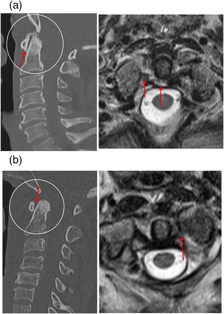
To identify trauma patients with confirmed tears of the transverse atlantal ligament on cervical MRI and measure several parameters of atlanto-axial alignment on cervical CT, including the anterior atlantodens interval, to determine which method is most sensitive in predicting transverse atlantal ligament injury.
Adult trauma patients who suffered a transverse atlantal ligament tear on cervical MRI were identified retrospectively. The cervical CT and MRI exams for these patients were reviewed for the following: anterior and lateral atlantodens interval widening, lateral C1 mass offset, C1-C2 rotatory subluxation, and transverse atlantal ligament injuries on cervical MRI.
Twenty-six patients were identified with a tear of the transverse atlantal ligament on cervical MRI. Twelve percent of these patients demonstrated an anterior dens interval measuring greater than 2 mm, 26% of patients demonstrated lateral mass offset of C1 on C2 (average offset of 2.4 mm), 18% of patients demonstrated an asymmetry greater than 1 mm between the left and right lateral atlantodens interval, and one patient demonstrated atlanto-axial rotation measuring greater than 20%. Ten patients had an accompanying C1 burst fracture and eight patients had a C2 fracture. One patient demonstrated widening of the atlanto-occipital joint space greater than 2 mm indicative of craniocervical dissociation injury.
An anterior atlantodens interval measuring greater than 2 mm is an unreliable methodology to screen trauma patients for transverse altantal ligament injuries and atlanto-axial instability. Moreover, C1 lateral mass offset, lateral atlantodens asymmetry, and atlanto-axial rotation were all poor predictors of transverse atlantal ligament tears.
回顾性横断面研究。
在颈椎磁共振成像(MRI)上识别确诊为寰椎横韧带撕裂的创伤患者,并在颈椎计算机断层扫描(CT)上测量寰枢椎对线的几个参数,包括寰齿前间隙,以确定哪种方法在预测寰椎横韧带损伤方面最敏感。
回顾性识别在颈椎MRI上发生寰椎横韧带撕裂的成年创伤患者。对这些患者的颈椎CT和MRI检查进行如下评估:寰齿前间隙和侧方间隙增宽、C1侧块偏移、C1-C2旋转半脱位以及颈椎MRI上的寰椎横韧带损伤情况。
在颈椎MRI上识别出26例寰椎横韧带撕裂患者。这些患者中,12%的患者寰齿前间隙测量值大于2mm,26%的患者表现出C1相对于C2的侧块偏移(平均偏移2.4mm),18%的患者左右侧寰齿侧方间隙不对称大于1mm,1例患者寰枢椎旋转大于20%。10例患者伴有C1爆裂骨折,8例患者有C2骨折。1例患者表现出寰枕关节间隙增宽大于2mm,提示颅颈分离损伤。
寰齿前间隙测量值大于2mm作为筛查创伤患者寰椎横韧带损伤和寰枢椎不稳是不可靠的方法。此外,C1侧块偏移、寰齿侧方不对称以及寰枢椎旋转均不能很好地预测寰椎横韧带撕裂。