Xu Xiaojun, Ruan Weiwei, Liu Fang, Gai Yongkang, Liu Qingyao, Su Ying, Liang Zhihou, Sun Xun, Lan Xiaoli
Department of Nuclear Medicine, Union Hospital, Tongji Medical College, Huazhong University of Science and Technology, Wuhan, China.
Hubei Province Key Laboratory of Molecular Imaging, Wuhan, China.
Front Aging Neurosci. 2022 Feb 10;13:789054. doi: 10.3389/fnagi.2021.789054. eCollection 2021.
F-APN-1607 is a novel tau positron emission tomography (PET) tracer characterized with high binding affinity for 3- and 4-repeat tau deposits. The aim was to analyze the spatial distribution of F-APN-1607 PET imaging in Alzheimer's disease (AD) subjects with different stages and to investigate the relationship between the change of tau deposition and overall disease progression.
We retrospectively analyzed the F-APN-1607 PET imaging of 31 subjects with clinically and imaging defined as AD. According to the Mini-Mental State Examination (MMSE) score, patients were divided into three groups, namely, mild (≥21, = 7), moderate (10-20, = 16), and severe (≤9, = 8). PET imaging was segmented to 70 regions of interest (ROIs) and extracted the standard uptake value (SUV) of each ROI. SUV ratio (SUVR) was calculated from the ratio of SUV in different brain regions to the cerebellar cortex. The regions were defined as positive and negative with unsupervised cluster analysis according to SUVR. The SUVRs of each region were compared among groups with the one-way ANOVA or Kruskal-Wallis test. Furthermore, the correlations between MMSE score and regional SUVR were calculated with Pearson or Spearman correlation analysis.
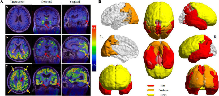
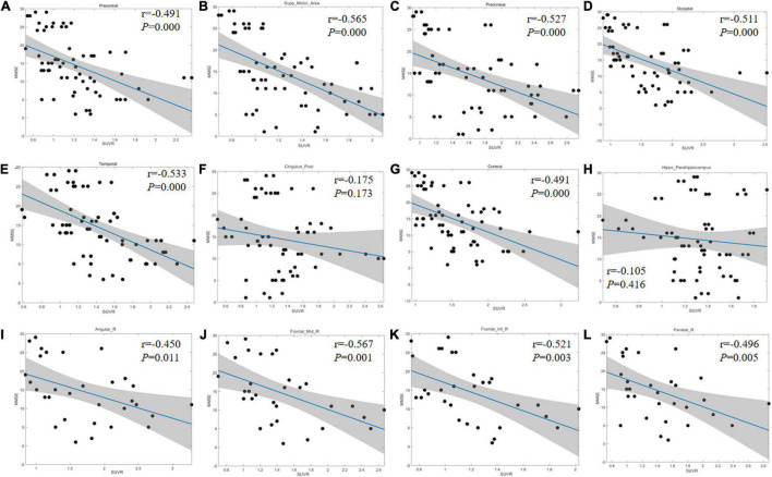
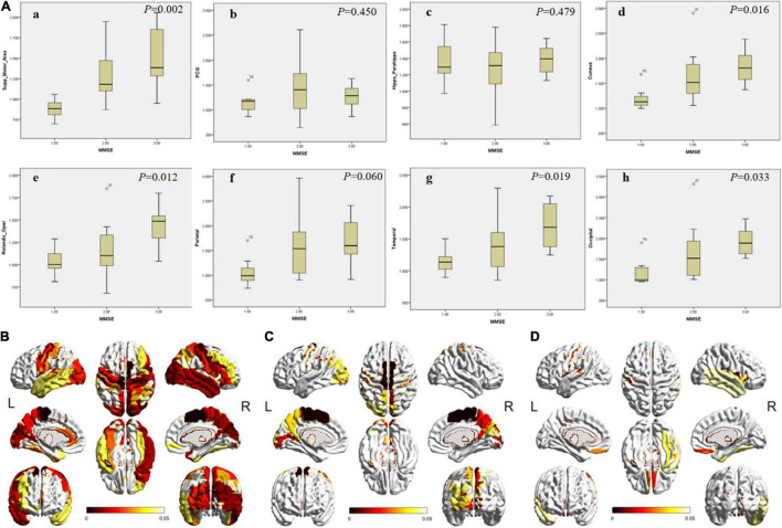
There were no significant differences among groups in gender (χ = 3.814, = 0.161), age of onset ( = 0.170), age ( = 0.109), and education level ( = 0.065). With the disease progression, the F-APN-1607 PET imaging showed the spread of tau deposition from the hippocampus, posterior cingulate gyrus (PCG), and lateral temporal cortex (LTC) to the parietal and occipital lobes, and finally to the frontal lobe. Between the mild and moderate groups, the main brain areas with significant differences in F-APN-1607 uptake were supplementary motor area (SMA), cuneus, precuneus, occipital lobule, paracentral lobule, right angular gyrus, and parietal, which could be used for early disease progression assessment ( < 0.05). There were significant differences in the frontal lobe, right temporal lobe, and fusiform gyrus between the moderate and severe groups, which might be suitable for the late-stage disease progression assessment ( < 0.05).
F-APN-1607 PET may serve as an effective imaging marker for visualizing the change pattern of tau protein deposition in AD patients, and its uptake level in certain brain regions is closely related to the severity of cognitive impairment. These indicate the potential of F-APN-1607 PET for the evaluation of the progression of AD.
F-APN-1607是一种新型的tau正电子发射断层扫描(PET)示踪剂,对3重复和4重复tau沉积物具有高结合亲和力。目的是分析F-APN-1607 PET成像在不同阶段阿尔茨海默病(AD)患者中的空间分布,并研究tau沉积变化与疾病整体进展之间的关系。
我们回顾性分析了31例临床和影像学诊断为AD的患者的F-APN-1607 PET成像。根据简易精神状态检查表(MMSE)评分,患者分为三组,即轻度(≥21,n = 7)、中度(10 - 20,n = 16)和重度(≤9,n = 8)。PET成像被分割为70个感兴趣区域(ROI),并提取每个ROI的标准摄取值(SUV)。SUV比率(SUVR)由不同脑区的SUV与小脑皮质的比率计算得出。根据SUVR通过无监督聚类分析将区域定义为阳性和阴性。使用单因素方差分析或Kruskal-Wallis检验比较各组间每个区域的SUVR。此外,通过Pearson或Spearman相关分析计算MMSE评分与区域SUVR之间的相关性。
各组在性别(χ² = 3.814,P = 0.161)、发病年龄(P = 0.170)、年龄(P = 0.109)和教育水平(P = 0.065)方面无显著差异。随着疾病进展,F-APN-1607 PET成像显示tau沉积从海马体、后扣带回皮质(PCG)和颞叶外侧皮质(LTC)扩散到顶叶和枕叶,最终扩散到额叶。在轻度和中度组之间,F-APN-1607摄取有显著差异的主要脑区是辅助运动区(SMA)、楔叶、楔前叶、枕叶小叶、中央旁小叶、右角回和顶叶,可用于疾病早期进展评估(P < 0.05)。在中度和重度组之间,额叶、右侧颞叶和梭状回有显著差异,可能适用于疾病晚期进展评估(P < 0.05)。
F-APN-1607 PET可能作为一种有效的成像标志物来可视化AD患者tau蛋白沉积的变化模式,其在某些脑区的摄取水平与认知障碍的严重程度密切相关。这些表明F-APN-1607 PET在评估AD进展方面的潜力。