Okabe Ryuichi, Ueki Yushi, Ohashi Riuko, Takeuchi Manabu, Hashimoto Satoru, Takahashi Takeshi, Shodo Ryusuke, Yamazaki Keisuke, Matsuyama Hiroshi, Umezu Hajime, Terai Shuji, Ajioka Yoichi, Horii Arata
Department of Otolaryngology-Head and Neck Surgery, Niigata University Graduate School of Medical and Dental Sciences, Niigata, Japan.
Department of Otorhinolaryngology, Nagaoka Red Cross Hospital, Niigata, Japan.
Front Surg. 2022 Feb 11;8:813260. doi: 10.3389/fsurg.2021.813260. eCollection 2021.
Early detection of head and neck carcinoma (HNC) as superficial HNC (SHNC) identified using recently developed optical techniques, such as magnifying endoscopy and narrow-band imaging (NBI), in combination with endoscopic surgeries enables minimally invasive treatment with favorable outcomes for HNC. This study aimed to identify the predictive factors for the rare but important clinical issue of SHNC, namely cervical lymph node metastasis (CLNM), following endoscopic resection.
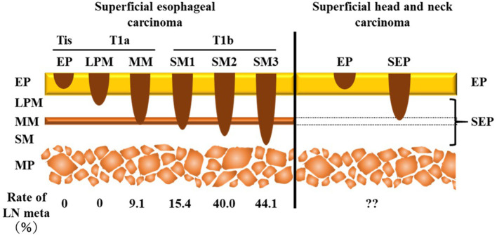
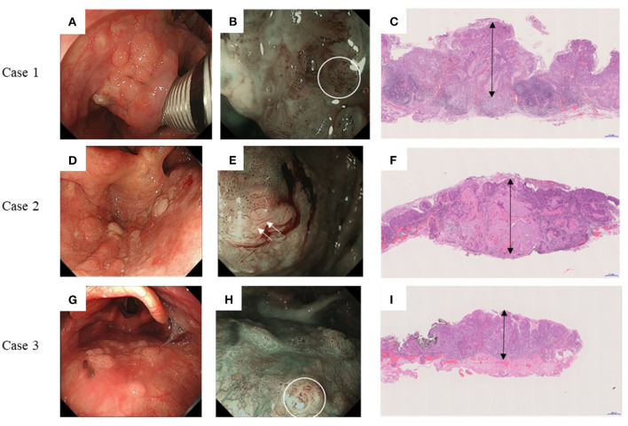
Sixty-nine patients with SHNC who underwent endoscopic resection were enrolled in the study. Clinical data, preoperative endoscopic findings, pathological findings, and treatment outcomes were retrospectively reviewed. Because the pharyngeal mucosa lacks the muscularis mucosa, we measured tumor thickness in permanent pathology as an alternative to the depth of invasion. Correlations with the occurrence of CLNM were statistically examined.
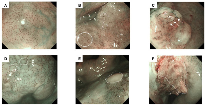
The 5-year disease-specific survival rate was 100%. Of 69 patients, 3 (4.3%) developed CLNM. All had subepithelial but not epithelial tumors. The 0-IIa type in the macroscopic findings, type B2/B3 vessels in narrow-band imaging, tumors ≥ pathological stage T2, lymphatic invasion, positive surgical margins, and tumor thickness >1,000 μm showed significant correlations with CLNM following endoscopic resection. Furthermore, the classification of type B vessels was significantly associated with tumor thickness.
The treatment outcomes following endoscopic resection for SHNC were favorable. The risk of CLNM following endoscopic resection in SHNC can be predicted by several preoperative endoscopic and postoperative pathological findings. Among them, the classification of type B vessels, which correlated with both tumor thickness and CLNM, might be a useful predictive factor.
利用最近开发的光学技术,如放大内镜和窄带成像(NBI),结合内镜手术,将头颈部癌(HNC)早期检测为浅表性HNC(SHNC),能够实现微创治疗,对头颈部癌有良好的治疗效果。本研究旨在确定内镜切除术后SHNC罕见但重要的临床问题——颈部淋巴结转移(CLNM)的预测因素。
69例接受内镜切除的SHNC患者纳入本研究。回顾性分析临床资料、术前内镜检查结果、病理检查结果及治疗效果。由于咽部黏膜缺乏黏膜肌层,我们在永久病理中测量肿瘤厚度,作为浸润深度的替代指标。对与CLNM发生的相关性进行统计学检验。
5年疾病特异性生存率为100%。69例患者中,3例(4.3%)发生CLNM。所有患者均为上皮下而非上皮肿瘤。内镜切除术后,宏观表现为0-IIa型、窄带成像中B2/B3型血管、肿瘤≥病理分期T2、有淋巴浸润、手术切缘阳性以及肿瘤厚度>1000μm与CLNM显著相关。此外,B型血管的分类与肿瘤厚度显著相关。
内镜切除治疗SHNC的效果良好。SHNC内镜切除术后CLNM的风险可通过术前内镜检查和术后病理检查结果进行预测。其中,与肿瘤厚度和CLNM均相关的B型血管分类可能是一个有用的预测因素。