• 文献检索
  • 文档翻译
  • 深度研究
  • 学术资讯
  • Suppr Zotero 插件Zotero 插件
  • 邀请有礼
  • 套餐&价格
  • 历史记录
应用&插件
Suppr Zotero 插件Zotero 插件浏览器插件Mac 客户端Windows 客户端微信小程序
定价
高级版会员购买积分包购买API积分包
服务
文献检索文档翻译深度研究API 文档MCP 服务
关于我们
关于 Suppr公司介绍联系我们用户协议隐私条款
关注我们

Suppr 超能文献

核心技术专利:CN118964589B侵权必究
粤ICP备2023148730 号-1Suppr @ 2026

文献检索

告别复杂PubMed语法,用中文像聊天一样搜索,搜遍4000万医学文献。AI智能推荐,让科研检索更轻松。

立即免费搜索

文件翻译

保留排版,准确专业,支持PDF/Word/PPT等文件格式,支持 12+语言互译。

免费翻译文档

深度研究

AI帮你快速写综述,25分钟生成高质量综述,智能提取关键信息,辅助科研写作。

立即免费体验

镰状细胞病患儿的视网膜和脉络膜厚度:一项横断面队列研究。

Retinal and choroidal thickness in pediatric patients with sickle cell disease: a cross-sectional cohort study.

作者信息

Prazeres Juliana, Lucatto Luiz Filipe, Ferreira Adriano, Moraes Nilva, Braga Josefina A P, Lima Luiz H, Regatieri Caio, Maia Maurício

机构信息

Department of Ophthalmology, Federal University of São Paulo, 806, Botucatu Street, São Paulo, 04026-062, Brazil.

Department of Pediatrics, Federal University of São Paulo, São Paulo, Brazil.

出版信息

Int J Retina Vitreous. 2022 Mar 4;8(1):15. doi: 10.1186/s40942-021-00351-3.

DOI:10.1186/s40942-021-00351-3
PMID:35246275
原文链接:https://pmc.ncbi.nlm.nih.gov/articles/PMC8895628/
Abstract

BACKGROUND

To measure the retinal/choroidal thicknesses in the macular area of asymptomatic pediatric patients with sickle cell disease (SCD).

METHODS

This cross-sectional cohort study included 40 children (79 eyes) with SCD and 19 control patients (36 eyes). All subjects underwent spectral-domain optical coherence tomography (SD-OCT) with enhanced-depth imaging OCT. Generalized Estimating Equations (GEE) were applied to compare the outcomes between groups. P ≤ 0.05 was considered significant.

RESULTS

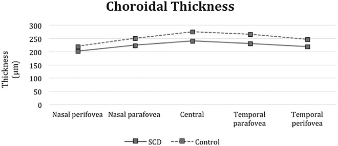
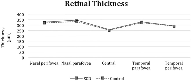
The choroidal thickness in the macular area in the study subfields was significantly thinner in the SCD eyes compared with control eyes (subfoveal subfield and temporal parafoveal subfield, p < 0.0001; nasal parafoveal subfield, p < 0.0001 temporal perifoveal subfield, p < 0.0001; and nasal perifoveal subfield, p < 0.0001). The variations in the retinal thickness were not significant.

CONCLUSION

EDI-OCT showed that the macular choroidal thickness is thinner in asymptomatic pediatric patients with SCD.

摘要

背景

测量无症状镰状细胞病(SCD)患儿黄斑区的视网膜/脉络膜厚度。

方法

这项横断面队列研究纳入了40例患有SCD的儿童(79只眼)和19例对照患者(36只眼)。所有受试者均接受了具有增强深度成像功能的光谱域光学相干断层扫描(SD-OCT)。采用广义估计方程(GEE)比较组间结果。P≤0.05被认为具有统计学意义。

结果

与对照眼相比,研究亚组中SCD眼黄斑区的脉络膜厚度明显更薄(中心凹下亚组和颞侧旁中心凹亚组,p<0.0001;鼻侧旁中心凹亚组,p<0.0001;颞侧中心凹周围亚组,p<0.0001;鼻侧中心凹周围亚组,p<0.0001)。视网膜厚度的差异不显著。

结论

增强深度成像光学相干断层扫描(EDI-OCT)显示,无症状SCD患儿的黄斑脉络膜厚度更薄。

https://cdn.ncbi.nlm.nih.gov/pmc/blobs/9424/8895628/3322c2a5964c/40942_2021_351_Fig3_HTML.jpg
https://cdn.ncbi.nlm.nih.gov/pmc/blobs/9424/8895628/512484e00023/40942_2021_351_Fig1_HTML.jpg
https://cdn.ncbi.nlm.nih.gov/pmc/blobs/9424/8895628/955ddadd28c0/40942_2021_351_Fig2_HTML.jpg
https://cdn.ncbi.nlm.nih.gov/pmc/blobs/9424/8895628/3322c2a5964c/40942_2021_351_Fig3_HTML.jpg
https://cdn.ncbi.nlm.nih.gov/pmc/blobs/9424/8895628/512484e00023/40942_2021_351_Fig1_HTML.jpg
https://cdn.ncbi.nlm.nih.gov/pmc/blobs/9424/8895628/955ddadd28c0/40942_2021_351_Fig2_HTML.jpg
https://cdn.ncbi.nlm.nih.gov/pmc/blobs/9424/8895628/3322c2a5964c/40942_2021_351_Fig3_HTML.jpg

相似文献

1
Retinal and choroidal thickness in pediatric patients with sickle cell disease: a cross-sectional cohort study.镰状细胞病患儿的视网膜和脉络膜厚度:一项横断面队列研究。
Int J Retina Vitreous. 2022 Mar 4;8(1):15. doi: 10.1186/s40942-021-00351-3.
2
Choroidal thinning as a new finding in Alzheimer's disease: evidence from enhanced depth imaging spectral domain optical coherence tomography.脉络膜变薄作为阿尔茨海默病的一项新发现:来自增强深度成像光谱域光学相干断层扫描的证据
J Alzheimers Dis. 2014;40(4):907-17. doi: 10.3233/JAD-132039.
3
Comparison of macular choroidal thickness in patients with pseudoexfoliation syndrome to normal control subjects with enhanced depth SD-OCT imaging.使用增强深度SD-OCT成像技术比较假性剥脱综合征患者与正常对照者的黄斑脉络膜厚度。
J Curr Ophthalmol. 2017 Jul 12;29(4):258-263. doi: 10.1016/j.joco.2017.06.009. eCollection 2017 Dec.
4
Spectral domain optical coherence tomography in patients with sickle cell disease.镰状细胞病患者的光谱域光学相干断层扫描
Br J Ophthalmol. 2015 Jul;99(7):967-72. doi: 10.1136/bjophthalmol-2014-305532. Epub 2015 Jan 16.
5
Retinal and choroidal thickness measurements using spectral domain optical coherence tomography in anterior and intermediate uveitis.应用频域光学相干断层扫描测量前葡萄膜炎和中间葡萄膜炎的视网膜和脉络膜厚度。
BMC Ophthalmol. 2014 Aug 30;14:103. doi: 10.1186/1471-2415-14-103.
6
Spectral-domain Optical Coherence Tomography Retinal and Choroidal Thickness Metric Repeatability in Age-related Macular Degeneration.光谱域光学相干断层扫描在年龄相关性黄斑变性中视网膜和脉络膜厚度测量指标的可重复性
Am J Ophthalmol. 2016 Jun;166:154-161. doi: 10.1016/j.ajo.2016.03.052. Epub 2016 Apr 9.
7
Macular and choroidal thicknesses in a healthy Hispanic population evaluated by high-definition spectral-domain optical coherence tomography (SD-OCT).通过高分辨率光谱域光学相干断层扫描(SD-OCT)评估健康西班牙裔人群的黄斑和脉络膜厚度。
Int J Retina Vitreous. 2020 Dec 7;6(1):66. doi: 10.1186/s40942-020-00270-9.
8
Evaluation of retinal nerve fiber layer and choroidal thickness with spectral domain optical coherence tomography in children with sickle cell anemia.利用频域光学相干断层扫描评估镰状细胞贫血患儿的视网膜神经纤维层和脉络膜厚度。
Turk J Pediatr. 2021;63(5):875-883. doi: 10.24953/turkjped.2021.05.015.
9
Repeatability of swept-source optical coherence tomography retinal and choroidal thickness measurements in neovascular age-related macular degeneration.扫频源光学相干断层扫描测量新生血管性年龄相关性黄斑变性视网膜和脉络膜厚度的可重复性
Br J Ophthalmol. 2017 May;101(5):603-608. doi: 10.1136/bjophthalmol-2016-308999. Epub 2016 Aug 4.
10
Macular choroidal thickness in patients with pseudoxanthoma elasticum measured by enhanced-depth imaging spectral-domain optical coherence tomography.应用增强深度成像谱域光学相干断层扫描测量弹性假黄瘤患者的脉络膜黄斑厚度。
Int Ophthalmol. 2020 Jul;40(7):1749-1758. doi: 10.1007/s10792-020-01343-2. Epub 2020 Mar 27.

引用本文的文献

1
Pitfalls of social media for aesthetic eye surgery patients: assessing YouTube's aesthetic canthoplasty content.社交媒体在眼部美容手术患者中的陷阱:评估 YouTube 上的美学重睑术内容。
Int Ophthalmol. 2024 Jun 25;44(1):279. doi: 10.1007/s10792-024-03197-4.

本文引用的文献

1
Retinal Thickness and Microvascular Changes in Children With Sickle Cell Disease Evaluated by Optical Coherence Tomography (OCT) and OCT Angiography.光学相干断层扫描(OCT)和 OCT 血管造影评估镰状细胞病儿童的视网膜厚度和微血管变化。
Am J Ophthalmol. 2020 Jan;209:88-98. doi: 10.1016/j.ajo.2019.08.019. Epub 2019 Aug 29.
2
Paramacular temporal atrophy in sickle cell disease occurs early in childhood.镰状细胞病患者的眶周颞部萎缩早在儿童期就会发生。
Br J Ophthalmol. 2019 Jul;103(7):906-910. doi: 10.1136/bjophthalmol-2018-312305. Epub 2018 Aug 21.
3
Optical Coherence Tomography Angiography and Ultra-widefield Fluorescein Angiography for Early Detection of Adolescent Sickle Retinopathy.
光学相干断层扫描血管造影术和超广角荧光素血管造影术用于青少年镰状视网膜病变的早期检测。
Am J Ophthalmol. 2017 Nov;183:91-98. doi: 10.1016/j.ajo.2017.08.010. Epub 2017 Aug 30.
4
Spectral domain optical coherence tomography in patients with sickle cell disease.镰状细胞病患者的光谱域光学相干断层扫描
Br J Ophthalmol. 2015 Jul;99(7):967-72. doi: 10.1136/bjophthalmol-2014-305532. Epub 2015 Jan 16.
5
Structural and functional correlation in sickle cell retinopathy using spectral-domain optical coherence tomography and scanning laser ophthalmoscope microperimetry.利用频域光相干断层扫描和扫描激光检眼镜微视野计研究镰状细胞性视网膜病变的结构和功能相关性。
Am J Ophthalmol. 2011 Oct;152(4):704-711.e2. doi: 10.1016/j.ajo.2011.03.035. Epub 2011 Jul 2.
6
Central macular splaying and outer retinal thinning in asymptomatic sickle cell patients by spectral-domain optical coherence tomography.频域光学相干断层扫描在无症状镰状细胞病患者中发现中心性黄斑分裂和外层视网膜变薄。
Am J Ophthalmol. 2011 Jun;151(6):990-994.e1. doi: 10.1016/j.ajo.2010.12.010. Epub 2011 Mar 31.
7
Temporal macular thinning on spectral-domain optical coherence tomography in proliferative sickle cell retinopathy.增殖性镰状细胞视网膜病变患者光谱域光学相干断层扫描显示颞侧黄斑变薄
Arch Ophthalmol. 2011 Feb;129(2):247-9. doi: 10.1001/archophthalmol.2010.357.
8
Sickle cell disease and the eye: old and new concepts.镰状细胞病与眼睛:旧概念与新概念。
Surv Ophthalmol. 2010 Jul-Aug;55(4):359-77. doi: 10.1016/j.survophthal.2009.11.004. Epub 2010 May 10.
9
Ocular manifestations of sickle cell disease.镰状细胞病的眼部表现。
Ann Trop Paediatr. 2010;30(1):19-26. doi: 10.1179/146532810X12637745451870.
10
Retinopathy in patients with sickle cell trait.镰状细胞性状患者的视网膜病变
Optometry. 2007 Nov;78(11):582-7. doi: 10.1016/j.optm.2007.04.100.