Amsterdam UMC, Location VUmc, Vrije Universiteit Amsterdam, Department of Anatomy and Neurosciences, Amsterdam Neuroscience, Amsterdam, The Netherlands.
Amsterdam UMC, location VUmc, Vrije Universiteit Amsterdam, Department of Pathology, Amsterdam Neuroscience, Amsterdam, The Netherlands.
Brain. 2022 Aug 27;145(8):2869-2881. doi: 10.1093/brain/awac093.
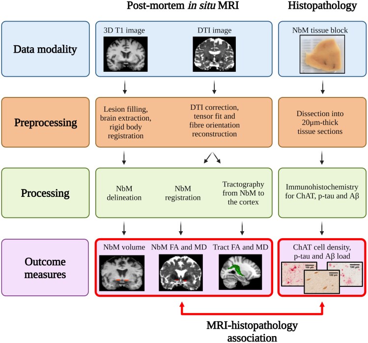
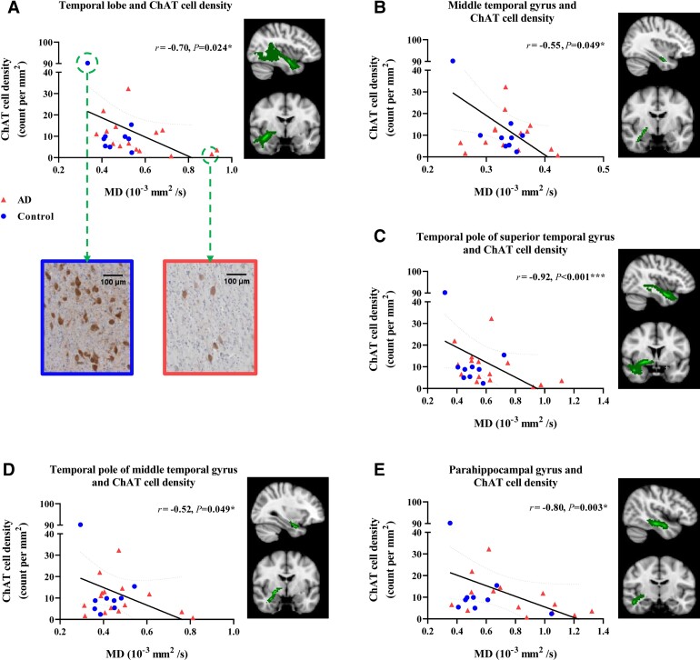
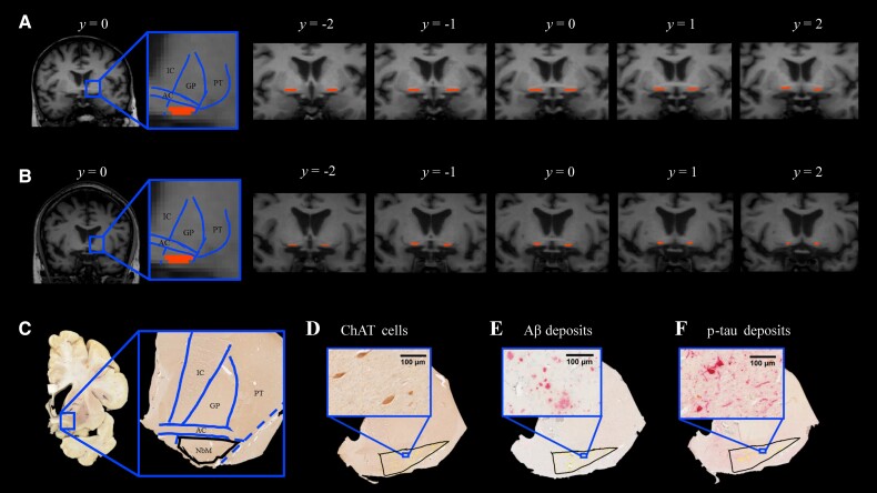
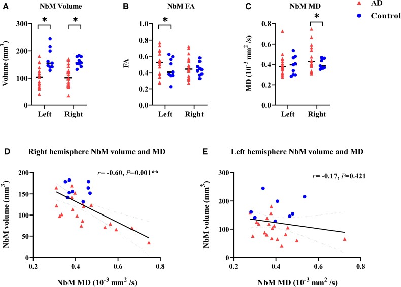
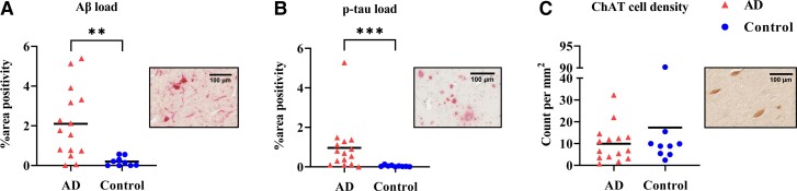
Cognitive deficits in Alzheimer's disease, specifically amnestic (memory dominant) deficits, are associated with cholinergic degeneration in the basal forebrain. The cholinergic nucleus within the basal forebrain, the nucleus basalis of Meynert, exhibits local atrophy and reduced cortical tract integrity on MRI, and reveals amyloid-β and phosphorylated-tau pathology at autopsy. To understand the pathophysiology of nucleus basalis of Meynert atrophy and its neocortical projections in Alzheimer's disease, we used a combined post-mortem in situ MRI and histopathology approach. A total of 19 Alzheimer's disease (10 amnestic and nine non-amnestic) and nine non-neurological control donors underwent 3 T T1-weighted MRI for anatomical delineation and volume assessment of the nucleus basalis of Meynert, and diffusion-weighted imaging for microstructural assessment of the nucleus and its projections. At subsequent brain autopsy, tissue dissection and immunohistochemistry were performed for amyloid-β, phosphorylated-tau and choline acetyltransferase. Compared to controls, we observed an MRI-derived volume reduction and altered microstructural integrity of the nucleus basalis of Meynert in Alzheimer's disease donors. Furthermore, decreased cholinergic cell density was associated with reduced integrity of the nucleus and its tracts to the temporal lobe, specifically to the temporal pole of the superior temporal gyrus, and the parahippocampal gyrus. Exploratory post hoc subgroup analyses indicated that cholinergic cell density could be associated with cortical tract alterations in amnestic Alzheimer's disease donors only. Our study illustrates that in Alzheimer's disease, cholinergic degeneration in the nucleus basalis of Meynert may contribute to damaged cortical projections, specifically to the temporal lobe, leading to cognitive deterioration.
阿尔茨海默病患者存在认知障碍,特别是健忘型(以记忆为主)认知障碍,与基底前脑胆碱能退化有关。基底前脑的胆碱能核团,即梅内尔特基底核,在 MRI 上表现为局部萎缩和皮质束完整性降低,尸检时显示淀粉样β和磷酸化 tau 病理学。为了了解梅内尔特基底核萎缩及其在阿尔茨海默病中的新皮质投射的病理生理学,我们采用了死后 MRI 与组织病理学相结合的方法。共有 19 名阿尔茨海默病(10 名健忘型和 9 名非健忘型)和 9 名非神经科对照供体接受了 3T T1 加权 MRI,用于解剖划分和梅内尔特基底核体积评估,以及弥散加权成像用于基底核及其投射的微观结构评估。在随后的脑尸检中,进行了组织解剖和免疫组织化学分析,以检测淀粉样β、磷酸化 tau 和胆碱乙酰转移酶。与对照组相比,我们在阿尔茨海默病供体中观察到 MRI 得出的基底核体积减少和微观结构完整性改变。此外,胆碱能细胞密度降低与基底核及其向颞叶,特别是向颞上回的颞极和海马旁回的束状结构完整性降低有关。探索性事后亚组分析表明,胆碱能细胞密度可能仅与健忘型阿尔茨海默病供体的皮质束改变有关。我们的研究表明,在阿尔茨海默病中,梅内尔特基底核的胆碱能退化可能导致皮质投射受损,特别是颞叶,导致认知恶化。