• 文献检索
  • 文档翻译
  • 深度研究
  • 学术资讯
  • Suppr Zotero 插件Zotero 插件
  • 邀请有礼
  • 套餐&价格
  • 历史记录
应用&插件
Suppr Zotero 插件Zotero 插件浏览器插件Mac 客户端Windows 客户端微信小程序
定价
高级版会员购买积分包购买API积分包
服务
文献检索文档翻译深度研究API 文档MCP 服务
关于我们
关于 Suppr公司介绍联系我们用户协议隐私条款
关注我们

Suppr 超能文献

核心技术专利:CN118964589B侵权必究
粤ICP备2023148730 号-1Suppr @ 2026

文献检索

告别复杂PubMed语法,用中文像聊天一样搜索,搜遍4000万医学文献。AI智能推荐,让科研检索更轻松。

立即免费搜索

文件翻译

保留排版,准确专业,支持PDF/Word/PPT等文件格式,支持 12+语言互译。

免费翻译文档

深度研究

AI帮你快速写综述,25分钟生成高质量综述,智能提取关键信息,辅助科研写作。

立即免费体验

用于胶质母细胞瘤、脑转移瘤和原发性中枢神经系统淋巴瘤鉴别诊断的术前血液检测。

Preoperative blood testing for glioblastoma, brain metastases, and primary central nervous system lymphoma differentiation.

作者信息

Wang Song-Quan, Yuan Qing, Zhang Guang-Tao, Qian Hai-Peng, Liu Zhi-Dan, Wang Jia-Wei, Cai Hong-Qing, Wan Jing-Hai

机构信息

Department of Neurosurgery, National Cancer Center/National Clinical Research Center for Cancer/Cancer Hospital, Chinese Academy of Medical Sciences and Peking Union Medical College, Beijing, China.

出版信息

Transl Cancer Res. 2022 Jan;11(1):63-71. doi: 10.21037/tcr-21-1957.

DOI:10.21037/tcr-21-1957
PMID:35261885
原文链接:https://pmc.ncbi.nlm.nih.gov/articles/PMC8841480/
Abstract

BACKGROUND

Differentiating glioblastoma (GBM), brain metastases, and primary central nervous system lymphoma (PCNSL) in clinical practice is difficult. This study aimed to evaluate the diagnostic value of routine blood biomarkers in patients with GBM, brain metastases, and PCNSL and find a preoperative differential diagnostic tool for these tumors.

METHODS

The perioperative medical records of 70 GBM, 41 PCNSL, and 81 brain metastases patients and their preoperative blood test results were compared and analyzed, and a diagnostic model to differentiate among them established.

RESULTS

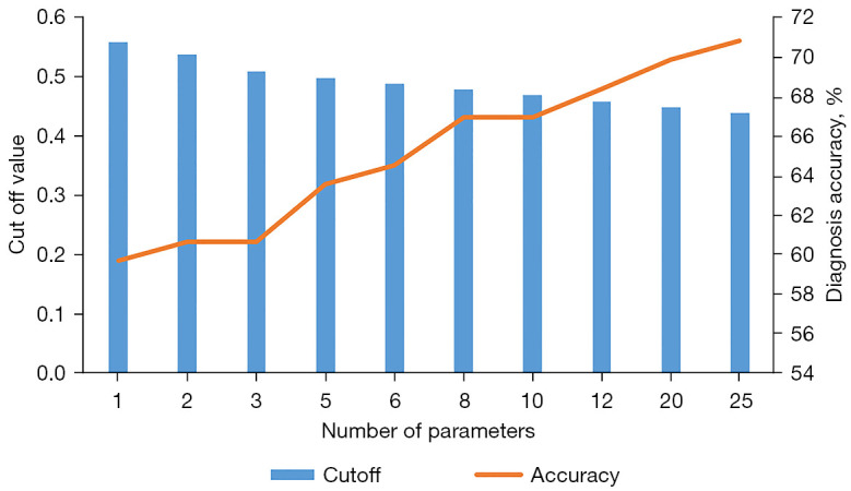
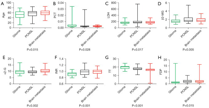
Patient age, plateletcrit, international normalized ratio (INR), and thrombin time (TT) were independently associated with differential diagnosis by multinomial logistic regression. Compared with GBM patients, brain metastases patients were significantly older (OR =1.055, 95% CI: 1.016-1.094, P=0.005) and had lower plateletcrit levels (OR =0.008, 95% CI: 0.004-0.017, P=0.027). In addition, patients with GBM had lower INR and higher TT than patients with the other two tumor types. A diagnostic model including these parameters, had an accuracy of 88.2% and 76.1% for brain metastases and GBM, respectively.

CONCLUSIONS

Preoperative plateletcrit, INR, and TT may be used as inexpensive blood diagnostic biomarkers for differentiating brain metastases from other intracranial malignant tumors.

摘要

背景

在临床实践中,鉴别胶质母细胞瘤(GBM)、脑转移瘤和原发性中枢神经系统淋巴瘤(PCNSL)具有挑战性。本研究旨在评估常规血液生物标志物在GBM、脑转移瘤和PCNSL患者中的诊断价值,并寻找这些肿瘤的术前鉴别诊断工具。

方法

比较并分析70例GBM、41例PCNSL和81例脑转移瘤患者的围手术期病历及其术前血液检查结果,并建立一个用于鉴别它们的诊断模型。

结果

通过多项逻辑回归分析,患者年龄、血小板压积、国际标准化比值(INR)和凝血酶时间(TT)与鉴别诊断独立相关。与GBM患者相比,脑转移瘤患者年龄显著更大(OR = 1.055,95%CI:1.016 - 1.094,P = 0.005)且血小板压积水平更低(OR = 0.008,95%CI:0.004 - 0.017,P = 0.027)。此外,GBM患者的INR低于其他两种肿瘤类型的患者,TT高于其他两种肿瘤类型的患者。包含这些参数的诊断模型对脑转移瘤和GBM的诊断准确率分别为88.2%和76.1%。

结论

术前血小板压积、INR和TT可作为廉价的血液诊断生物标志物,用于鉴别脑转移瘤与其他颅内恶性肿瘤。

https://cdn.ncbi.nlm.nih.gov/pmc/blobs/9e21/8841480/edf854721c00/tcr-11-01-63-f2.jpg
https://cdn.ncbi.nlm.nih.gov/pmc/blobs/9e21/8841480/3aab62870391/tcr-11-01-63-f1.jpg
https://cdn.ncbi.nlm.nih.gov/pmc/blobs/9e21/8841480/edf854721c00/tcr-11-01-63-f2.jpg
https://cdn.ncbi.nlm.nih.gov/pmc/blobs/9e21/8841480/3aab62870391/tcr-11-01-63-f1.jpg
https://cdn.ncbi.nlm.nih.gov/pmc/blobs/9e21/8841480/edf854721c00/tcr-11-01-63-f2.jpg

相似文献

1
Preoperative blood testing for glioblastoma, brain metastases, and primary central nervous system lymphoma differentiation.用于胶质母细胞瘤、脑转移瘤和原发性中枢神经系统淋巴瘤鉴别诊断的术前血液检测。
Transl Cancer Res. 2022 Jan;11(1):63-71. doi: 10.21037/tcr-21-1957.
2
What is the Best Preoperative Quantitative Indicator to Differentiate Primary Central Nervous System Lymphoma from Glioblastoma?鉴别原发性中枢神经系统淋巴瘤与胶质母细胞瘤的最佳术前定量指标是什么?
World Neurosurg. 2023 Apr;172:e517-e523. doi: 10.1016/j.wneu.2023.01.065. Epub 2023 Jan 21.
3
Intraoperative Flow Cytometry Enables the Differentiation of Primary Central Nervous System Lymphoma from Glioblastoma.术中流式细胞术有助于鉴别原发性中枢神经系统淋巴瘤和胶质母细胞瘤。
World Neurosurg. 2018 Apr;112:e261-e268. doi: 10.1016/j.wneu.2018.01.033. Epub 2018 Jan 9.
4
Machine learning applications for the differentiation of primary central nervous system lymphoma from glioblastoma on imaging: a systematic review and meta-analysis.基于影像的机器学习应用于原发性中枢神经系统淋巴瘤与胶质母细胞瘤的鉴别:系统评价和荟萃分析。
Neurosurg Focus. 2018 Nov 1;45(5):E5. doi: 10.3171/2018.8.FOCUS18325.
5
Primary central nervous system lymphoma and atypical glioblastoma: differentiation using the initial area under the curve derived from dynamic contrast-enhanced MR and the apparent diffusion coefficient.原发中枢神经系统淋巴瘤和非典型性脑胶质瘤:基于动态对比增强磁共振的初始曲线下面积和表观扩散系数的鉴别诊断。
Eur Radiol. 2017 Apr;27(4):1344-1351. doi: 10.1007/s00330-016-4484-2. Epub 2016 Jul 19.
6
Can morphological MRI differentiate between primary central nervous system lymphoma and glioblastoma?形态学磁共振成像能否区分原发性中枢神经系统淋巴瘤和胶质母细胞瘤?
Cancer Imaging. 2016 Nov 29;16(1):40. doi: 10.1186/s40644-016-0098-9.
7
Differentiating between Primary Central Nervous System Lymphoma and Glioblastoma: The Diagnostic Value of Combining F-fluorodeoxyglucose Positron Emission Tomography with Arterial Spin Labeling.原发性中枢神经系统淋巴瘤与胶质母细胞瘤的鉴别:结合 F-氟脱氧葡萄糖正电子发射断层扫描与动脉自旋标记的诊断价值。
Neurol Med Chir (Tokyo). 2021 Jun 15;61(6):367-375. doi: 10.2176/nmc.oa.2020-0375. Epub 2021 May 8.
8
Differentiation between primary CNS lymphoma and glioblastoma: qualitative and quantitative analysis using arterial spin labeling MR imaging.原发中枢神经系统淋巴瘤与胶质母细胞瘤的鉴别诊断:动脉自旋标记 MRI 的定性和定量分析。
Eur Radiol. 2018 Sep;28(9):3801-3810. doi: 10.1007/s00330-018-5359-5. Epub 2018 Apr 4.
9
Differentiating between Glioblastoma and Primary CNS Lymphoma Using Combined Whole-tumor Histogram Analysis of the Normalized Cerebral Blood Volume and the Apparent Diffusion Coefficient.利用归一化脑血容量和表观扩散系数的全肿瘤直方图分析鉴别胶质母细胞瘤和原发性中枢神经系统淋巴瘤。
Magn Reson Med Sci. 2019 Jan 10;18(1):53-61. doi: 10.2463/mrms.mp.2017-0135. Epub 2018 May 31.
10
Perfusion Weighted MR Imaging may Differentiate Primary CNS Lymphoma from other Homogeneously Enhancing Brain Tumors.
Neuroradiol J. 2008 Dec 17;21(5):637-44. doi: 10.1177/197140090802100505.

引用本文的文献

1
Predictors for the Differentiation between Glioblastoma, Primary Central Nervous System Lymphoma, and Metastasis in Patients with a Solitary Enhancing Intracranial Mass.孤立性颅内强化肿块患者中胶质母细胞瘤、原发性中枢神经系统淋巴瘤和转移瘤鉴别的预测因素
Asian J Neurosurg. 2024 Jun 6;19(2):186-201. doi: 10.1055/s-0044-1787051. eCollection 2024 Jun.
2
Prognostic Values of Systemic Inflammatory Immunological Markers in Glioblastoma: A Systematic Review and Meta-Analysis.胶质母细胞瘤中全身炎症免疫标志物的预后价值:一项系统评价和荟萃分析
Cancers (Basel). 2023 Jun 25;15(13):3339. doi: 10.3390/cancers15133339.
3
Artificial neural network-based diagnostic models for lung cancer combining conventional indicators with tumor markers.

本文引用的文献

1
Preoperative plateletcrit is a Prognostic Biomarker for Survival in Patients with Non-Small Cell Lung Cancer.术前血小板比容是预测非小细胞肺癌患者生存的生物标志物。
J Cancer. 2020 Feb 25;11(10):2800-2807. doi: 10.7150/jca.41122. eCollection 2020.
2
Differentiation of primary central nervous system lymphoma from high-grade glioma and brain metastasis using arterial spin labeling and dynamic contrast-enhanced magnetic resonance imaging.利用动脉自旋标记和动态对比增强磁共振成像对原发性中枢神经系统淋巴瘤与高级别胶质瘤和脑转移瘤进行鉴别。
Eur J Radiol. 2019 Mar;112:59-64. doi: 10.1016/j.ejrad.2019.01.008. Epub 2019 Jan 8.
3
基于人工神经网络的结合常规指标与肿瘤标志物的肺癌诊断模型。
Exp Biol Med (Maywood). 2023 May;248(10):829-838. doi: 10.1177/15353702231177013. Epub 2023 Jul 4.
4
Systemic immune-inflammation landscape in brain metastasis needing neurosurgical resection: Analysis of 230 consecutive cases in a single center.脑转移瘤行神经外科切除术患者的系统性免疫炎症特征:单中心 230 例连续病例分析。
Immun Inflamm Dis. 2022 Oct;10(10):e694. doi: 10.1002/iid3.694.
Brain metastases.
脑转移瘤。
Nat Rev Dis Primers. 2019 Jan 17;5(1):5. doi: 10.1038/s41572-018-0055-y.
4
MRI as a diagnostic biomarker for differentiating primary central nervous system lymphoma from glioblastoma: A systematic review and meta-analysis.MRI 作为一种诊断生物标志物,用于区分原发性中枢神经系统淋巴瘤与胶质母细胞瘤:系统评价和荟萃分析。
J Magn Reson Imaging. 2019 Aug;50(2):560-572. doi: 10.1002/jmri.26602. Epub 2019 Jan 14.
5
Epidemiology and Overview of Gliomas.胶质瘤的流行病学与概述
Semin Oncol Nurs. 2018 Dec;34(5):420-429. doi: 10.1016/j.soncn.2018.10.001. Epub 2018 Nov 2.
6
Significance and Prognostic Value of The Coagulation Profile in Patients with Glioblastoma: Implications for Personalized Therapy.胶质母细胞瘤患者凝血指标的意义及预后价值:对个性化治疗的启示
World Neurosurg. 2019 Jan;121:e621-e629. doi: 10.1016/j.wneu.2018.09.177. Epub 2018 Oct 3.
7
Perfusion Computed Tomography Parameters Are Useful for Differentiating Glioblastoma, Lymphoma, and Metastasis.灌注计算机断层扫描参数有助于鉴别胶质母细胞瘤、淋巴瘤和转移瘤。
World Neurosurg. 2018 Nov;119:e890-e897. doi: 10.1016/j.wneu.2018.07.291. Epub 2018 Aug 9.
8
Diagnostic value of preoperative inflammatory markers in patients with glioma: a multicenter cohort study.术前炎症标志物对胶质瘤患者的诊断价值:一项多中心队列研究。
J Neurosurg. 2018 Sep;129(3):583-592. doi: 10.3171/2017.3.JNS161648. Epub 2017 Nov 3.
9
Primary central nervous system lymphoma.原发性中枢神经系统淋巴瘤。
Crit Rev Oncol Hematol. 2017 May;113:97-110. doi: 10.1016/j.critrevonc.2017.03.019. Epub 2017 Mar 18.
10
Evaluation of platelet indices as a useful marker in papillary thyroid carcinoma.血小板指标作为甲状腺乳头状癌有用标志物的评估
Bratisl Lek Listy. 2017;118(3):153-155. doi: 10.4149/BLL_2017_030.