• 文献检索
  • 文档翻译
  • 深度研究
  • 学术资讯
  • Suppr Zotero 插件Zotero 插件
  • 邀请有礼
  • 套餐&价格
  • 历史记录
应用&插件
Suppr Zotero 插件Zotero 插件浏览器插件Mac 客户端Windows 客户端微信小程序
定价
高级版会员购买积分包购买API积分包
服务
文献检索文档翻译深度研究API 文档MCP 服务
关于我们
关于 Suppr公司介绍联系我们用户协议隐私条款
关注我们

Suppr 超能文献

核心技术专利:CN118964589B侵权必究
粤ICP备2023148730 号-1Suppr @ 2026

文献检索

告别复杂PubMed语法,用中文像聊天一样搜索,搜遍4000万医学文献。AI智能推荐,让科研检索更轻松。

立即免费搜索

文件翻译

保留排版,准确专业,支持PDF/Word/PPT等文件格式,支持 12+语言互译。

免费翻译文档

深度研究

AI帮你快速写综述,25分钟生成高质量综述,智能提取关键信息,辅助科研写作。

立即免费体验

孤立性颅内强化肿块患者中胶质母细胞瘤、原发性中枢神经系统淋巴瘤和转移瘤鉴别的预测因素

Predictors for the Differentiation between Glioblastoma, Primary Central Nervous System Lymphoma, and Metastasis in Patients with a Solitary Enhancing Intracranial Mass.

作者信息

Chuthip Pornthida, Sitthinamsuwan Bunpot, Witthiwej Theerapol, Tansirisithikul Chottiwat, Khumpalikit Inthira, Nunta-Aree Sarun

机构信息

Division of Neurosurgery, Department of Surgery, Faculty of Medicine Siriraj Hospital, Mahidol University, Bangkok, Thailand.

Department of Surgery, Pattani Hospital, Pattani, Thailand.

出版信息

Asian J Neurosurg. 2024 Jun 6;19(2):186-201. doi: 10.1055/s-0044-1787051. eCollection 2024 Jun.

DOI:10.1055/s-0044-1787051
PMID:38974428
原文链接:https://pmc.ncbi.nlm.nih.gov/articles/PMC11226298/
Abstract

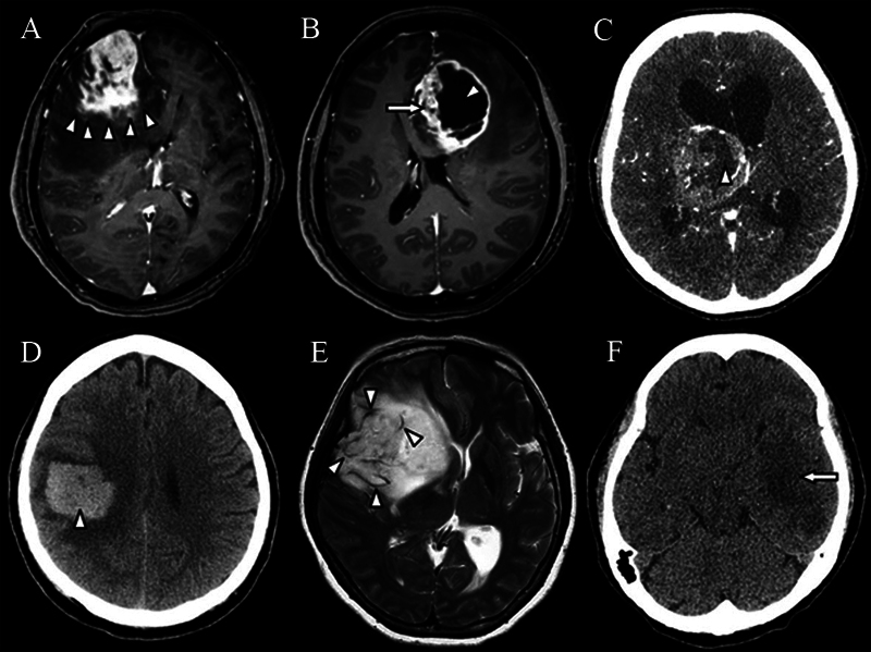
Differentiation between glioblastoma (GBM), primary central nervous system lymphoma (PCNSL), and metastasis is important in decision-making before surgery. However, these malignant brain tumors have overlapping features. This study aimed to identify predictors differentiating between GBM, PCNSL, and metastasis.  Patients with a solitary intracranial enhancing tumor and a histopathological diagnosis of GBM, PCNSL, or metastasis were investigated. All patients with intracranial lymphoma had PCNSL without extracranial involvement. Demographic, clinical, and radiographic data were analyzed to determine their associations with the tumor types.  The predictors associated with GBM were functional impairment (  = 0.001), large tumor size (  < 0.001), irregular tumor margin (  < 0.001), heterogeneous contrast enhancement (  < 0.001), central necrosis (  < 0.001), intratumoral hemorrhage (  = 0.018), abnormal flow void (  < 0.001), and hypodensity component on noncontrast cranial computed tomography (CT) scan (  < 0.001). The predictors associated with PCNSL comprised functional impairment (  = 0.005), deep-seated tumor location (  = 0.006), homogeneous contrast enhancement (  < 0.001), absence of cystic appearance (  = 0.008), presence of hypointensity component on precontrast cranial T1-weighted magnetic resonance imaging (MRI;  = 0.027), and presence of isodensity component on noncontrast cranial CT (  < 0.008). Finally, the predictors for metastasis were an infratentorial (  < 0.001) or extra-axial tumor location (  = 0.035), smooth tumor margin (  < 0.001), and presence of isointensity component on cranial fluid-attenuated inversion recovery MRI (  = 0.047).  These predictors may be used to differentiate between GBM, PCNSL, and metastasis, and they are useful in clinical management.

摘要

在手术前的决策过程中,区分胶质母细胞瘤(GBM)、原发性中枢神经系统淋巴瘤(PCNSL)和脑转移瘤非常重要。然而,这些恶性脑肿瘤具有一些重叠的特征。本研究旨在确定区分GBM、PCNSL和脑转移瘤的预测因素。

对患有孤立性颅内强化肿瘤且组织病理学诊断为GBM、PCNSL或脑转移瘤的患者进行了调查。所有颅内淋巴瘤患者均为无颅外受累的PCNSL。分析了人口统计学、临床和影像学数据,以确定它们与肿瘤类型的关联。

与GBM相关的预测因素包括功能障碍(P = 0.001)、肿瘤体积大(P < 0.001)、肿瘤边缘不规则(P < 0.001)、不均匀对比增强(P < 0.001)、中央坏死(P < 0.001)、瘤内出血(P = 0.018)、异常血流空洞(P < 0.001)以及非增强头颅计算机断层扫描(CT)上的低密度成分(P < 0.001)。与PCNSL相关的预测因素包括功能障碍(P = 0.005)、肿瘤位于深部(P = 0.006)、均匀对比增强(P < 0.001)、无囊状表现(P = 0.008)、在头颅T1加权磁共振成像(MRI)平扫前存在低信号成分(P = 0.027)以及在非增强头颅CT上存在等密度成分(P < 0.008)。最后,脑转移瘤的预测因素为幕下(P < 0.001)或轴外肿瘤位置(P = 0.035)、肿瘤边缘光滑(P < 0.001)以及在头颅液体衰减反转恢复序列MRI上存在等信号成分(P = 0.047)。

这些预测因素可用于区分GBM、PCNSL和脑转移瘤,对临床管理具有重要意义。

https://cdn.ncbi.nlm.nih.gov/pmc/blobs/6bca/11226298/df0f9223facd/10-1055-s-0044-1787051-i2360035-3.jpg
https://cdn.ncbi.nlm.nih.gov/pmc/blobs/6bca/11226298/1e4a30f1c606/10-1055-s-0044-1787051-i2360035-1.jpg
https://cdn.ncbi.nlm.nih.gov/pmc/blobs/6bca/11226298/023f7d14ce25/10-1055-s-0044-1787051-i2360035-2.jpg
https://cdn.ncbi.nlm.nih.gov/pmc/blobs/6bca/11226298/df0f9223facd/10-1055-s-0044-1787051-i2360035-3.jpg

相似文献

1
Predictors for the Differentiation between Glioblastoma, Primary Central Nervous System Lymphoma, and Metastasis in Patients with a Solitary Enhancing Intracranial Mass.孤立性颅内强化肿块患者中胶质母细胞瘤、原发性中枢神经系统淋巴瘤和转移瘤鉴别的预测因素
Asian J Neurosurg. 2024 Jun 6;19(2):186-201. doi: 10.1055/s-0044-1787051. eCollection 2024 Jun.
2
Can morphological MRI differentiate between primary central nervous system lymphoma and glioblastoma?形态学磁共振成像能否区分原发性中枢神经系统淋巴瘤和胶质母细胞瘤?
Cancer Imaging. 2016 Nov 29;16(1):40. doi: 10.1186/s40644-016-0098-9.
3
Primary central nervous system lymphoma and glioblastoma differentiation based on conventional magnetic resonance imaging by high-throughput SIFT features.基于高通量SIFT特征的常规磁共振成像对原发性中枢神经系统淋巴瘤和胶质母细胞瘤的鉴别诊断
Int J Neurosci. 2018 Jul;128(7):608-618. doi: 10.1080/00207454.2017.1408613. Epub 2017 Dec 12.
4
Primary central nervous system lymphoma and atypical glioblastoma: differentiation using the initial area under the curve derived from dynamic contrast-enhanced MR and the apparent diffusion coefficient.原发中枢神经系统淋巴瘤和非典型性脑胶质瘤:基于动态对比增强磁共振的初始曲线下面积和表观扩散系数的鉴别诊断。
Eur Radiol. 2017 Apr;27(4):1344-1351. doi: 10.1007/s00330-016-4484-2. Epub 2016 Jul 19.
5
F-FDG PET/CT in immunocompetent patients with primary central nervous system lymphoma: Differentiation from glioblastoma and correlation with DWI.免疫功能正常的原发性中枢神经系统淋巴瘤患者的 F-FDG PET/CT:与胶质母细胞瘤的鉴别诊断及与 DWI 的相关性。
Eur J Radiol. 2018 Jul;104:26-32. doi: 10.1016/j.ejrad.2018.04.020. Epub 2018 Apr 22.
6
What is the Best Preoperative Quantitative Indicator to Differentiate Primary Central Nervous System Lymphoma from Glioblastoma?鉴别原发性中枢神经系统淋巴瘤与胶质母细胞瘤的最佳术前定量指标是什么?
World Neurosurg. 2023 Apr;172:e517-e523. doi: 10.1016/j.wneu.2023.01.065. Epub 2023 Jan 21.
7
Intraoperative Flow Cytometry Enables the Differentiation of Primary Central Nervous System Lymphoma from Glioblastoma.术中流式细胞术有助于鉴别原发性中枢神经系统淋巴瘤和胶质母细胞瘤。
World Neurosurg. 2018 Apr;112:e261-e268. doi: 10.1016/j.wneu.2018.01.033. Epub 2018 Jan 9.
8
Differentiating Glioblastoma from Primary Central Nervous System Lymphoma: The Value of Shaping and Nonenhancing Peritumoral Hyperintense Gyral Lesion on FLAIR Imaging.FLAIR 成像上形态学及非增强性瘤周高信号脑回样病变对鉴别胶质母细胞瘤与原发性中枢神经系统淋巴瘤的价值。
World Neurosurg. 2021 May;149:e696-e704. doi: 10.1016/j.wneu.2021.01.114. Epub 2021 Feb 3.
9
Atypical primary central nervous system lymphoma and glioblastoma: multiparametric differentiation based on non-enhancing volume, apparent diffusion coefficient, and arterial spin labeling.非增强容积、表观弥散系数和动脉自旋标记在原发性中枢神经系统淋巴瘤和胶质母细胞瘤中的多参数鉴别诊断
Eur Radiol. 2023 Aug;33(8):5357-5367. doi: 10.1007/s00330-023-09681-2. Epub 2023 May 12.
10
Machine learning applications for the differentiation of primary central nervous system lymphoma from glioblastoma on imaging: a systematic review and meta-analysis.基于影像的机器学习应用于原发性中枢神经系统淋巴瘤与胶质母细胞瘤的鉴别:系统评价和荟萃分析。
Neurosurg Focus. 2018 Nov 1;45(5):E5. doi: 10.3171/2018.8.FOCUS18325.

本文引用的文献

1
A Deep Learning Model for Preoperative Differentiation of Glioblastoma, Brain Metastasis and Primary Central Nervous System Lymphoma: A Pilot Study.一种用于胶质母细胞瘤、脑转移瘤和原发性中枢神经系统淋巴瘤术前鉴别诊断的深度学习模型:一项初步研究。
Front Oncol. 2022 Feb 24;12:816638. doi: 10.3389/fonc.2022.816638. eCollection 2022.
2
Preoperative blood testing for glioblastoma, brain metastases, and primary central nervous system lymphoma differentiation.用于胶质母细胞瘤、脑转移瘤和原发性中枢神经系统淋巴瘤鉴别诊断的术前血液检测。
Transl Cancer Res. 2022 Jan;11(1):63-71. doi: 10.21037/tcr-21-1957.
3
Diagnostic efficacy of apparent diffusion coefficient measurements in differentiation of malignant intra-axial brain tumors.
表观扩散系数测量在鉴别恶性脑内肿瘤中的诊断效能。
Turk J Med Sci. 2021 Feb 26;51(1):256-267. doi: 10.3906/sag-2006-1.
4
Utility of Percentage Signal Recovery and Baseline Signal in DSC-MRI Optimized for Relative CBV Measurement for Differentiating Glioblastoma, Lymphoma, Metastasis, and Meningioma.用于相对 CBV 测量的 DSC-MRI 的百分比信号恢复和基线信号的效用,可用于鉴别胶质母细胞瘤、淋巴瘤、转移瘤和脑膜瘤。
AJNR Am J Neuroradiol. 2019 Sep;40(9):1445-1450. doi: 10.3174/ajnr.A6153. Epub 2019 Aug 1.
5
Machine learning for semi-automated classification of glioblastoma, brain metastasis and central nervous system lymphoma using magnetic resonance advanced imaging.利用磁共振高级成像技术进行胶质母细胞瘤、脑转移瘤和中枢神经系统淋巴瘤半自动分类的机器学习
Ann Transl Med. 2019 Jun;7(11):232. doi: 10.21037/atm.2018.08.05.
6
Primary CNS Lymphoma in the Elderly: The Challenge.老年人原发性中枢神经系统淋巴瘤:挑战。
Acta Haematol. 2019;141(3):138-145. doi: 10.1159/000495284. Epub 2019 Feb 15.
7
Differentiation of primary central nervous system lymphoma from high-grade glioma and brain metastasis using arterial spin labeling and dynamic contrast-enhanced magnetic resonance imaging.利用动脉自旋标记和动态对比增强磁共振成像对原发性中枢神经系统淋巴瘤与高级别胶质瘤和脑转移瘤进行鉴别。
Eur J Radiol. 2019 Mar;112:59-64. doi: 10.1016/j.ejrad.2019.01.008. Epub 2019 Jan 8.
8
Perfusion Computed Tomography Parameters Are Useful for Differentiating Glioblastoma, Lymphoma, and Metastasis.灌注计算机断层扫描参数有助于鉴别胶质母细胞瘤、淋巴瘤和转移瘤。
World Neurosurg. 2018 Nov;119:e890-e897. doi: 10.1016/j.wneu.2018.07.291. Epub 2018 Aug 9.
9
Primary brain tumours in adults.成人原发性脑肿瘤。
Lancet. 2018 Aug 4;392(10145):432-446. doi: 10.1016/S0140-6736(18)30990-5. Epub 2018 Jul 27.
10
F-FDG PET/CT in immunocompetent patients with primary central nervous system lymphoma: Differentiation from glioblastoma and correlation with DWI.免疫功能正常的原发性中枢神经系统淋巴瘤患者的 F-FDG PET/CT:与胶质母细胞瘤的鉴别诊断及与 DWI 的相关性。
Eur J Radiol. 2018 Jul;104:26-32. doi: 10.1016/j.ejrad.2018.04.020. Epub 2018 Apr 22.