• 文献检索
  • 文档翻译
  • 深度研究
  • 学术资讯
  • Suppr Zotero 插件Zotero 插件
  • 邀请有礼
  • 套餐&价格
  • 历史记录
应用&插件
Suppr Zotero 插件Zotero 插件浏览器插件Mac 客户端Windows 客户端微信小程序
定价
高级版会员购买积分包购买API积分包
服务
文献检索文档翻译深度研究API 文档MCP 服务
关于我们
关于 Suppr公司介绍联系我们用户协议隐私条款
关注我们

Suppr 超能文献

核心技术专利:CN118964589B侵权必究
粤ICP备2023148730 号-1Suppr @ 2026

文献检索

告别复杂PubMed语法,用中文像聊天一样搜索,搜遍4000万医学文献。AI智能推荐,让科研检索更轻松。

立即免费搜索

文件翻译

保留排版,准确专业,支持PDF/Word/PPT等文件格式,支持 12+语言互译。

免费翻译文档

深度研究

AI帮你快速写综述,25分钟生成高质量综述,智能提取关键信息,辅助科研写作。

立即免费体验

子宫内膜癌:动态对比增强 MRI 示踪动力学模型在术前风险评估中的应用。

Endometrial carcinoma: use of tracer kinetic modeling of dynamic contrast-enhanced MRI for preoperative risk assessment.

机构信息

Department of Radiology, Key Laboratory of Birth Defects and Related Diseases of Women and Children (Sichuan University), Ministry of Education, West China Second University Hospital, Sichuan University, No.20, Section 3, Renmin South Road, Chengdu, 610041, Sichuan, China.

Department of Oncologic Imaging, National Cancer Center, Singapore, 169610, Singapore.

出版信息

Cancer Imaging. 2022 Mar 9;22(1):14. doi: 10.1186/s40644-022-00452-8.

DOI:10.1186/s40644-022-00452-8
PMID:35264244
原文链接:https://pmc.ncbi.nlm.nih.gov/articles/PMC8908697/
Abstract

BACKGROUND

To compare two tracer kinetic models in predicting of preoperative risk types in endometrial carcinoma (EC) using DCE-MRI.

METHODS

A prospective study of patients with EC was conducted with institutional ethics approval and written informed consent. DCE-MRI data was analyzed using the extended Tofts (ET) and the distributed parameter (DP) models. DCE parameters blood flow (F), mean transit time, blood volume (Vp), extravascular extracellular volume (Ve), permeability surface area product (PS), extraction fraction, transfer constant (Ktrans), and efflux rate (Kep) between high- and low-risk EC were compared using the Mann-Whitney test. Bland-Altman analysis was utilized to compare parameter consistency and Spearman test to assess parameter correlation. Diagnostic performance of DCE parameters was analyzed by receiver-operating characteristic curve and compared with traditional MRI assessment.

RESULTS

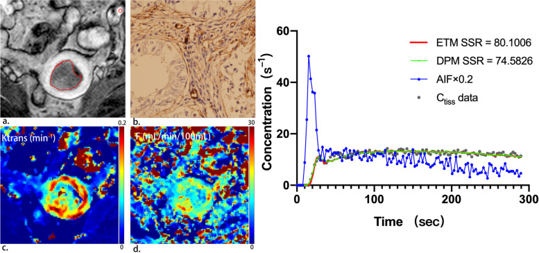
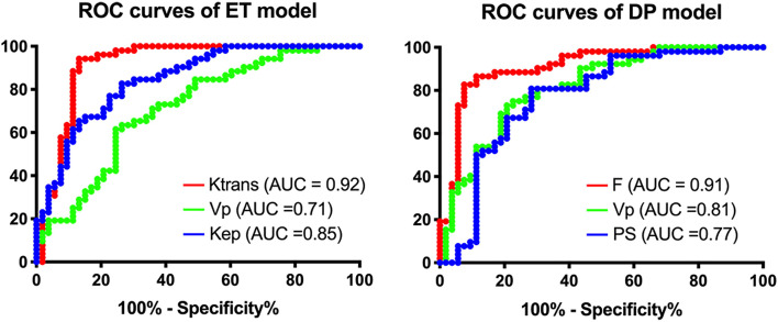
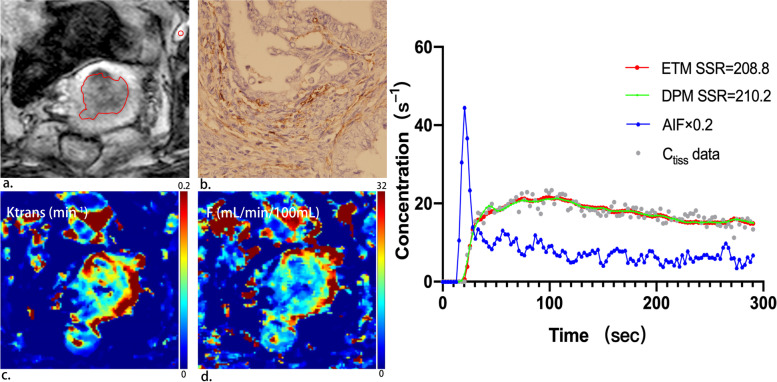
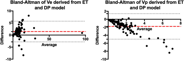
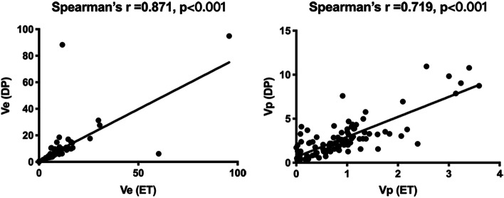
Fifty-one patients comprised the study group. Patients with high-risk EC exhibited significantly lower Ktrans, Kep, F, Vp and PS (P < 0.001). ET-derived Ktrans and DP-derived F attained AUC of 0.92 and 0.91, respectively. Bland-Altman analysis showed that the consistency of Ve or Vp between the two models was low (P < 0.001) while Spearman test showed a strong correlation (r = 0.719, 0.871). Both Ktrans and F showed higher accuracy in predicting EC risk types than traditional MRI assessment.

CONCLUSIONS

Kinetic parameters derived from DCE-MRI revealed a more hypovascular microenvironment for high risk EC than to low- risk ones, providing potential imaging biomarkers in preoperative risk assessment that might improve individualized surgical planning and management of EC.

摘要

背景

比较两种示踪剂动力学模型在使用 DCE-MRI 预测子宫内膜癌(EC)术前风险类型中的应用。

方法

本研究经机构伦理审查委员会批准,并获得患者书面知情同意,前瞻性纳入 EC 患者。采用扩展 Tofts(ET)和分布参数(DP)模型分析 DCE-MRI 数据。使用 Mann-Whitney 检验比较高、低危 EC 之间的 DCE 参数(F、平均通过时间、血容量(Vp)、血管外细胞外容积(Ve)、渗透率表面积乘积(PS)、提取分数、转移常数(Ktrans)和流出率(Kep)。Bland-Altman 分析用于比较参数一致性,Spearman 检验用于评估参数相关性。通过接收者操作特征曲线分析 DCE 参数的诊断性能,并与传统 MRI 评估进行比较。

结果

本研究共纳入 51 例患者。高危 EC 患者的 Ktrans、Kep、F、Vp 和 PS 明显降低(P<0.001)。ET 衍生的 Ktrans 和 DP 衍生的 F 的 AUC 分别为 0.92 和 0.91。Bland-Altman 分析表明两种模型之间 Ve 或 Vp 的一致性较差(P<0.001),Spearman 检验表明相关性较强(r=0.719,0.871)。与传统 MRI 评估相比,Ktrans 和 F 对预测 EC 风险类型的准确性更高。

结论

DCE-MRI 衍生的动力学参数显示高危 EC 具有更低的血管生成微环境,为术前风险评估提供了潜在的影像学生物标志物,可能改善 EC 的个体化手术规划和管理。

https://cdn.ncbi.nlm.nih.gov/pmc/blobs/e4c8/8908697/c9ce49a77077/40644_2022_452_Fig5_HTML.jpg
https://cdn.ncbi.nlm.nih.gov/pmc/blobs/e4c8/8908697/de272f1af951/40644_2022_452_Fig1_HTML.jpg
https://cdn.ncbi.nlm.nih.gov/pmc/blobs/e4c8/8908697/a24c00a4937a/40644_2022_452_Fig2_HTML.jpg
https://cdn.ncbi.nlm.nih.gov/pmc/blobs/e4c8/8908697/77721a8bdb6d/40644_2022_452_Fig3_HTML.jpg
https://cdn.ncbi.nlm.nih.gov/pmc/blobs/e4c8/8908697/e659f77c22e1/40644_2022_452_Fig4_HTML.jpg
https://cdn.ncbi.nlm.nih.gov/pmc/blobs/e4c8/8908697/c9ce49a77077/40644_2022_452_Fig5_HTML.jpg

相似文献

1
Endometrial carcinoma: use of tracer kinetic modeling of dynamic contrast-enhanced MRI for preoperative risk assessment.子宫内膜癌:动态对比增强 MRI 示踪动力学模型在术前风险评估中的应用。
Cancer Imaging. 2022 Mar 9;22(1):14. doi: 10.1186/s40644-022-00452-8.
2
Dynamic contrast-enhanced MRI in endometrial carcinoma identifies patients at increased risk of recurrence.动态对比增强磁共振成像在子宫内膜癌中可识别出复发风险增加的患者。
Eur Radiol. 2013 Oct;23(10):2916-25. doi: 10.1007/s00330-013-2901-3. Epub 2013 Jun 4.
3
Application of Distributed Parameter Model to Assessment of Glioma IDH Mutation Status by Dynamic Contrast-Enhanced Magnetic Resonance Imaging.应用分布参数模型评估动态对比增强磁共振成像中的脑胶质瘤 IDH 突变状态。
Contrast Media Mol Imaging. 2020 Nov 22;2020:8843084. doi: 10.1155/2020/8843084. eCollection 2020.
4
Dual-input two-compartment pharmacokinetic model of dynamic contrast-enhanced magnetic resonance imaging in hepatocellular carcinoma.肝细胞癌动态对比增强磁共振成像的双输入双室药代动力学模型
World J Gastroenterol. 2016 Apr 7;22(13):3652-62. doi: 10.3748/wjg.v22.i13.3652.
5
Dependence of DCE-MRI biomarker values on analysis algorithm.动态对比增强磁共振成像(DCE-MRI)生物标志物值对分析算法的依赖性。
PLoS One. 2015 Jul 24;10(7):e0130168. doi: 10.1371/journal.pone.0130168. eCollection 2015.
6
On the potential use of dynamic contrast-enhanced (DCE) MRI parameters as radiomic features of cervical cancer.探讨动态对比增强(DCE)MRI 参数作为宫颈癌放射组学特征的潜在应用。
Med Phys. 2019 Nov;46(11):5098-5109. doi: 10.1002/mp.13821. Epub 2019 Sep 26.
7
Quantitative dynamic contrast-enhanced parameters and intravoxel incoherent motion facilitate the prediction of TP53 status and risk stratification of early-stage endometrial carcinoma.定量动态对比增强参数和体素内不相干运动有助于预测早期子宫内膜癌的 TP53 状态和风险分层。
Radiol Oncol. 2023 Jun 21;57(2):257-269. doi: 10.2478/raon-2023-0023. eCollection 2023 Jun 1.
8
Differentiation of grade II and III oligodendrogliomas from grade II and III astrocytomas: a histogram analysis of perfusion parameters derived from dynamic contrast-enhanced (DCE) and dynamic susceptibility contrast (DSC) MRI.II级和III级少突胶质细胞瘤与II级和III级星形细胞瘤的鉴别:基于动态对比增强(DCE)和动态磁敏感对比(DSC)MRI获得的灌注参数的直方图分析
Acta Radiol. 2018 Jun;59(6):723-731. doi: 10.1177/0284185117728981. Epub 2017 Sep 1.
9
Use of biexponential and stretched exponential models of intravoxel incoherent motion and dynamic contrast-enhanced magnetic resonance imaging to assess the proliferation of endometrial carcinoma.使用体素内不相干运动的双指数和拉伸指数模型以及动态对比增强磁共振成像评估子宫内膜癌的增殖情况。
Quant Imaging Med Surg. 2023 Apr 1;13(4):2568-2581. doi: 10.21037/qims-22-688. Epub 2023 Feb 16.
10
Differentiation of Benign from Malignant Adnexal Masses by Dynamic Contrast-Enhanced MRI (DCE-MRI): Quantitative and Semi-quantitative analysis at 3-Tesla MRI.通过动态对比增强磁共振成像(DCE-MRI)鉴别良性与恶性附件肿块:3特斯拉磁共振成像的定量和半定量分析
Asian Pac J Cancer Prev. 2019 Apr 29;20(4):1073-1079. doi: 10.31557/APJCP.2019.20.4.1073.

引用本文的文献

1
Preoperative risk assessment of endometrial cancer using histogram analysis of weighted and quantitative MRI images.使用加权和定量MRI图像的直方图分析对子宫内膜癌进行术前风险评估。
Abdom Radiol (NY). 2025 Jun 27. doi: 10.1007/s00261-025-05069-6.
2
Dynamic contrast‑enhanced MRI combined with intravoxel incoherent motion in quantitative evaluation for preoperative risk stratification of resectable rectal adenocarcinoma.动态对比增强磁共振成像联合体素内不相干运动在可切除直肠腺癌术前风险分层定量评估中的应用
Oncol Lett. 2024 Nov 20;29(2):68. doi: 10.3892/ol.2024.14814. eCollection 2025 Feb.
3
Co-reactivity pattern of glucose metabolism and blood perfusion revealing DNA mismatch repair deficiency based on PET/DCE-MRI in endometrial cancer.

本文引用的文献

1
Quantitative diffusion and perfusion MRI in the evaluation of endometrial cancer: validation with histopathological parameters.定量扩散和灌注 MRI 在子宫内膜癌评估中的应用:与组织病理学参数的验证。
Br J Radiol. 2021 Sep 1;94(1125):20210054. doi: 10.1259/bjr.20210054. Epub 2021 Jun 16.
2
Differentiation of endometrial adenocarcinoma from adenocarcinoma of cervix using kinetic parameters derived from DCE-MRI.使用 DCE-MRI 得出的动力学参数区分子宫内膜腺癌和宫颈癌腺癌。
Eur J Radiol. 2020 Sep;130:109190. doi: 10.1016/j.ejrad.2020.109190. Epub 2020 Jul 24.
3
Cancer statistics, 2020.
基于 PET/DCE-MRI 的子宫内膜癌葡萄糖代谢与血供的共反应性模式揭示 DNA 错配修复缺陷。
Cancer Imaging. 2024 Nov 25;24(1):161. doi: 10.1186/s40644-024-00805-5.
4
Applying dynamic contrast-enhanced MRI tracer kinetic models to differentiate benign and malignant soft tissue tumors.应用动态对比增强 MRI 示踪剂动力学模型鉴别良恶性软组织肿瘤。
Cancer Imaging. 2024 May 21;24(1):64. doi: 10.1186/s40644-024-00710-x.
5
Evaluation of multi-parameter MRI in preoperative staging of endometrial carcinoma.多参数磁共振成像在子宫内膜癌术前分期中的评估
Eur J Radiol Open. 2024 Mar 23;12:100559. doi: 10.1016/j.ejro.2024.100559. eCollection 2024 Jun.
6
Quantitative dynamic contrast-enhanced parameters and intravoxel incoherent motion facilitate the prediction of TP53 status and risk stratification of early-stage endometrial carcinoma.定量动态对比增强参数和体素内不相干运动有助于预测早期子宫内膜癌的 TP53 状态和风险分层。
Radiol Oncol. 2023 Jun 21;57(2):257-269. doi: 10.2478/raon-2023-0023. eCollection 2023 Jun 1.
7
Dynamic contrast-enhanced MR imaging of uterine endometrial carcinoma with/without squamous differentiation.有/无鳞状分化的子宫内膜癌的动态对比增强磁共振成像
Abdom Radiol (NY). 2023 Aug;48(8):2494-2502. doi: 10.1007/s00261-023-03934-w. Epub 2023 May 8.
8
Use of biexponential and stretched exponential models of intravoxel incoherent motion and dynamic contrast-enhanced magnetic resonance imaging to assess the proliferation of endometrial carcinoma.使用体素内不相干运动的双指数和拉伸指数模型以及动态对比增强磁共振成像评估子宫内膜癌的增殖情况。
Quant Imaging Med Surg. 2023 Apr 1;13(4):2568-2581. doi: 10.21037/qims-22-688. Epub 2023 Feb 16.
9
Tissue Characteristics of Endometrial Carcinoma Analyzed by Quantitative Synthetic MRI and Diffusion-Weighted Imaging.定量合成磁共振成像和扩散加权成像分析子宫内膜癌的组织特征
Diagnostics (Basel). 2022 Nov 25;12(12):2956. doi: 10.3390/diagnostics12122956.
10
Quantitative texture analysis based on dynamic contrast enhanced MRI for differential diagnosis between primary thymic lymphoma from thymic carcinoma.基于动态对比增强 MRI 的定量纹理分析在原发性胸腺瘤与胸腺癌鉴别诊断中的应用。
Sci Rep. 2022 Jul 24;12(1):12629. doi: 10.1038/s41598-022-16393-y.
癌症统计数据,2020 年。
CA Cancer J Clin. 2020 Jan;70(1):7-30. doi: 10.3322/caac.21590. Epub 2020 Jan 8.
4
Nanoparticles' interactions with vasculature in diseases.纳米粒子在疾病中与血管的相互作用。
Chem Soc Rev. 2019 Oct 28;48(21):5381-5407. doi: 10.1039/c9cs00309f.
5
Poor outcome in hypoxic endometrial carcinoma is related to vascular density.缺氧型子宫内膜癌预后不良与血管密度相关。
Br J Cancer. 2019 May;120(11):1037-1044. doi: 10.1038/s41416-019-0461-2. Epub 2019 Apr 23.
6
Can preoperative magnetic resonance imaging replace intraoperative frozen sectioning in the evaluation of myometrial invasion for early-stage endometrial carcinoma?在早期子宫内膜癌肌层浸润评估中,术前磁共振成像能否取代术中冰冻切片检查?
Ginekol Pol. 2019;90(3):128-133. doi: 10.5603/GP.2019.0023.
7
A Prospective Observational Study Evaluating the Accuracy of MRI in Predicting the Extent of Disease in Endometrial Cancer.一项评估MRI预测子宫内膜癌疾病范围准确性的前瞻性观察性研究。
Indian J Surg Oncol. 2019 Mar;10(1):220-224. doi: 10.1007/s13193-018-0832-9. Epub 2018 Dec 17.
8
Pathologic Prognostic Factors in Endometrial Carcinoma (Other Than Tumor Type and Grade).子宫内膜癌的病理预后因素(肿瘤类型和分级除外)
Int J Gynecol Pathol. 2019 Jan;38 Suppl 1(Iss 1 Suppl 1):S93-S113. doi: 10.1097/PGP.0000000000000524.
9
Cancer incidence and mortality patterns in Europe: Estimates for 40 countries and 25 major cancers in 2018.欧洲癌症发病率和死亡率模式:2018 年 40 个国家和 25 种主要癌症的估计值。
Eur J Cancer. 2018 Nov;103:356-387. doi: 10.1016/j.ejca.2018.07.005. Epub 2018 Aug 9.
10
Preoperative Evaluation of Myometrial Invasion in Endometrial Carcinoma: Prospective Intra-individual Comparison of Magnetic Resonance Volumetry, Diffusion-weighted and Dynamic Contrast-enhanced Magnetic Resonance Imaging.子宫内膜癌肌层浸润的术前评估:磁共振容积测定法、扩散加权及动态对比增强磁共振成像的前瞻性个体内比较
Anticancer Res. 2018 Aug;38(8):4813-4817. doi: 10.21873/anticanres.12791.