Cai Jingyao, Lyu Xing, Huang Peiying, Li Shisheng, Chen Ruohong, Chen Zhiyang, Sun Mei, Zeng Ling, Wu Fengxi, Hu Min
Department of Laboratory Medicine, The Second Xiangya Hospital of Central South University, Changsha, China.
Department of Otolaryngology-Head and Neck Surgery, The Second Xiangya Hospital, Central South University, Changsha, China.
Front Med (Lausanne). 2022 Feb 25;9:854570. doi: 10.3389/fmed.2022.854570. eCollection 2022.
Obstructive sleep apnea-hypopnea syndrome (OSA) may cause liver fibrosis, and liver fibrosis serum biomarkers plays an important role on the diagnosis of liver fibrosis. In addition, this study aimed to observe the changes of 4 serum markers and Chitinase 3-like protein 1 (CHII3L1) levels in OSA patients with different disease severity and explore their interactions. And then, we examined whether intermittent hypoxia (IH) exposure can activate hepatic stellate cell.
74 OSA patients in Second Xiangya hospital from January 2021 to October 2021 was selected and categorized into mild, moderate, and severe groups according to AHI. In addition, 20 subjects were selected as the control group. Serum levels of liver fibrosis markers were determined by electrochemiluminescence immunoassay. Hepatic stellate cells were exposed to intermittent IH or normoxia (RA). Results were analyzed using the SPSS software.
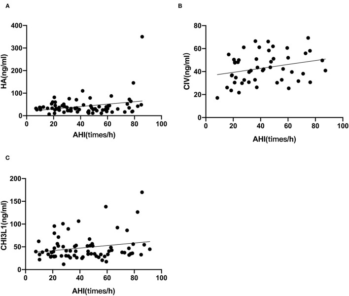
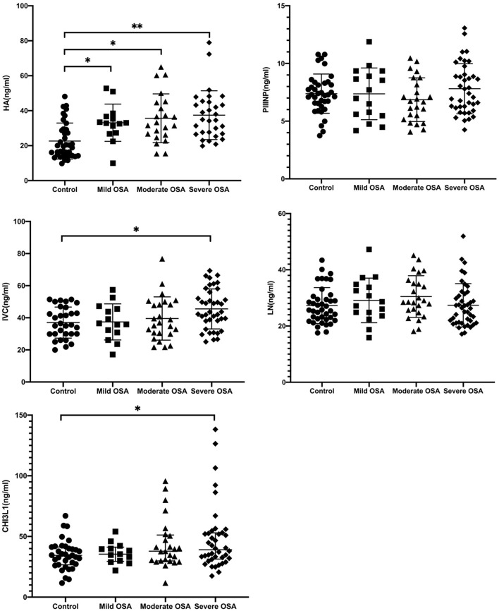
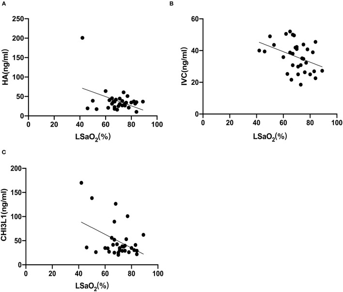
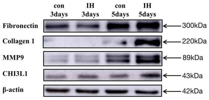
There was a significant increase in serum hyaluronic acid (HA), collagen type IV (CIV) and CHI3L1 levels in OSA patients compared with control group. Specifically, serum liver fibrosis markers HA, CIV and CHI3L1 levels were positively correlated with apnea-hypopnea index (AHI), but negatively correlated with the lowest saturation oxygen (LSaO) respectively. The LX-2 cells (human hepatic stellate cell line) exposed to IH showed significant increases in fibrotic protein expression.
OSA might either directly or indirectly trigger or exacerbate liver fibrosis, possibly via IH-related pathways.
阻塞性睡眠呼吸暂停低通气综合征(OSA)可能导致肝纤维化,而肝纤维化血清生物标志物在肝纤维化诊断中起重要作用。此外,本研究旨在观察不同疾病严重程度的OSA患者4种血清标志物和几丁质酶3样蛋白1(CHII3L1)水平的变化,并探讨它们之间的相互作用。然后,我们研究了间歇性缺氧(IH)暴露是否能激活肝星状细胞。
选取2021年1月至2021年10月在湘雅二医院就诊的74例OSA患者,根据呼吸暂停低通气指数(AHI)分为轻度、中度和重度组。另外,选取20名受试者作为对照组。采用电化学发光免疫分析法测定血清肝纤维化标志物水平。将肝星状细胞暴露于间歇性IH或常氧(RA)环境中。使用SPSS软件分析结果。
与对照组相比,OSA患者血清透明质酸(HA)、IV型胶原(CIV)和CHI3L1水平显著升高。具体而言血清肝纤维化标志物HA、CIV和CHI3L1水平与呼吸暂停低通气指数(AHI)呈正相关,但分别与最低血氧饱和度(LSaO)呈负相关。暴露于IH的LX-2细胞(人肝星状细胞系)纤维化蛋白表达显著增加。
OSA可能直接或间接触发或加重肝纤维化,可能通过与IH相关的途径。