• 文献检索
  • 文档翻译
  • 深度研究
  • 学术资讯
  • Suppr Zotero 插件Zotero 插件
  • 邀请有礼
  • 套餐&价格
  • 历史记录
应用&插件
Suppr Zotero 插件Zotero 插件浏览器插件Mac 客户端Windows 客户端微信小程序
定价
高级版会员购买积分包购买API积分包
服务
文献检索文档翻译深度研究API 文档MCP 服务
关于我们
关于 Suppr公司介绍联系我们用户协议隐私条款
关注我们

Suppr 超能文献

核心技术专利:CN118964589B侵权必究
粤ICP备2023148730 号-1Suppr @ 2026

文献检索

告别复杂PubMed语法,用中文像聊天一样搜索,搜遍4000万医学文献。AI智能推荐,让科研检索更轻松。

立即免费搜索

文件翻译

保留排版,准确专业,支持PDF/Word/PPT等文件格式,支持 12+语言互译。

免费翻译文档

深度研究

AI帮你快速写综述,25分钟生成高质量综述,智能提取关键信息,辅助科研写作。

立即免费体验

犬股动脉狭窄模型中狭窄后血流紊乱的定量评估:矢量血流成像的早期经验

Quantitative Evaluation of Post-stenotic Blood Flow Disturbance in Canine Femoral Artery Stenosis Model: An Early Experience With Vector Flow Imaging.

作者信息

Zhao Rui, Zheng Haining, Wang Wei, Du Yigang, Tong Yisha, Wen Chaoyang

机构信息

Department of Ultrasound, Peking University International Hospital, Beijing, China.

Department of Ultrasound, The Fourth Medical Center of PLA General Hospital, Beijing, China.

出版信息

Front Cardiovasc Med. 2022 Feb 24;9:829825. doi: 10.3389/fcvm.2022.829825. eCollection 2022.

DOI:10.3389/fcvm.2022.829825
PMID:35282375
原文链接:https://pmc.ncbi.nlm.nih.gov/articles/PMC8907590/
Abstract

OBJECTIVE

To investigate the value of Vector Flow Imaging (V Flow) in the assessment of post-stenotic turbulence in the canine arterial stenosis model.

MATERIALS AND METHODS

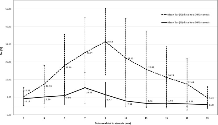
Canine femoral artery stenosis models were established using ameroid constrictors in 12 beagle dogs. 50% and then 70% femoral artery stenoses were confirmed by selective femoral artery angiography. V Flow was used to measure femoral artery flow turbulence index (Tur) preoperatively as a baseline. After establishing of a 50% and then 70% stenoses, the Tur indices were recorded in the femoral artery at 1, 3, 5, 7, 9, 11, 13, 15, 17, and 19 mm distal to the stenosis.

RESULTS

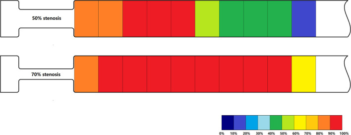
Baseline Tur indices of normal canine femoral arteries were <1% in 11 of 12 cases (91.7%). Distal to a 50% stenosis, the Tur index (>1%) was recorded in 83.3-100% cases between 1 and 9 mm, 41.7-58.3% between 11 and 17 mm, and 16.7% at 19 mm. For a 70% stenosis, the Tur index (>1%) occurred in 81.8-100% cases between 1 and 17 mm distal to the stenosis, and 63.6% at 19 mm. The Tur index peaked around 7 mm or 2.3 times of the initial vessel diameter (3 mm) downstream for a 50% stenosis and 11 mm or 3.7 times of vessel diameter downstream for a 70% stenosis.

CONCLUSION

V Flow with Tur index measurement adds quantitative information of post-stenotic turbulence when assessing an arterial stenosis with ultrasound. Tur index of 1% seems a useful threshold for assessment of flow turbulence in this small sample study. Further studies with larger sample size are needed to evaluate the value of V Flow in clinical applications.

摘要

目的

探讨矢量血流成像(V Flow)在评估犬动脉狭窄模型中狭窄后湍流情况的价值。

材料与方法

使用阿梅氏缩窄器在12只比格犬中建立犬股动脉狭窄模型。通过选择性股动脉血管造影确认股动脉狭窄程度分别为50%和70%。术前使用V Flow测量股动脉血流湍流指数(Tur)作为基线值。在建立50%和70%狭窄后,记录狭窄远端1、3、5、7、9、11、13、15、17和19毫米处股动脉的Tur指数。

结果

12例正常犬股动脉的基线Tur指数中,11例(91.7%)<1%。在50%狭窄远端,1至9毫米处83.3% - 100%的病例记录到Tur指数(>1%),11至17毫米处为41.7% - 58.3%,19毫米处为16.7%。对于70%狭窄,狭窄远端1至17毫米处81.8% - 100%的病例出现Tur指数(>1%),19毫米处为63.6%。对于50%狭窄,Tur指数在狭窄下游约7毫米或初始血管直径(3毫米)的2.3倍处达到峰值;对于70%狭窄,Tur指数在狭窄下游11毫米或血管直径的3.7倍处达到峰值。

结论

在超声评估动脉狭窄时,V Flow结合Tur指数测量可增加狭窄后湍流的定量信息。在本小样本研究中,1%的Tur指数似乎是评估血流湍流的有用阈值。需要进一步进行更大样本量的研究来评估V Flow在临床应用中的价值。

https://cdn.ncbi.nlm.nih.gov/pmc/blobs/3f8d/8907590/5524f37a97c9/fcvm-09-829825-g0007.jpg
https://cdn.ncbi.nlm.nih.gov/pmc/blobs/3f8d/8907590/92175ce047ee/fcvm-09-829825-g0001.jpg
https://cdn.ncbi.nlm.nih.gov/pmc/blobs/3f8d/8907590/8d45040b080a/fcvm-09-829825-g0002.jpg
https://cdn.ncbi.nlm.nih.gov/pmc/blobs/3f8d/8907590/c7094a05f906/fcvm-09-829825-g0003.jpg
https://cdn.ncbi.nlm.nih.gov/pmc/blobs/3f8d/8907590/bf189907e0c8/fcvm-09-829825-g0004.jpg
https://cdn.ncbi.nlm.nih.gov/pmc/blobs/3f8d/8907590/f9ed7c91301f/fcvm-09-829825-g0005.jpg
https://cdn.ncbi.nlm.nih.gov/pmc/blobs/3f8d/8907590/49824f066909/fcvm-09-829825-g0006.jpg
https://cdn.ncbi.nlm.nih.gov/pmc/blobs/3f8d/8907590/5524f37a97c9/fcvm-09-829825-g0007.jpg

相似文献

1
Quantitative Evaluation of Post-stenotic Blood Flow Disturbance in Canine Femoral Artery Stenosis Model: An Early Experience With Vector Flow Imaging.犬股动脉狭窄模型中狭窄后血流紊乱的定量评估:矢量血流成像的早期经验
Front Cardiovasc Med. 2022 Feb 24;9:829825. doi: 10.3389/fcvm.2022.829825. eCollection 2022.
2
Corrigendum: Quantitative evaluation of post-stenotic blood flow disturbance in canine femoral artery stenosis model: An early experience with vector flow imaging.勘误:犬股动脉狭窄模型中狭窄后血流紊乱的定量评估:矢量流成像的早期经验。
Front Cardiovasc Med. 2022 Jul 20;9:979992. doi: 10.3389/fcvm.2022.979992. eCollection 2022.
3
Evaluation of Turbulence Index and Flow Pattern for Atherosclerotic Carotid Stenosis: A High-Frame-Rate Vector Flow Imaging Study.评价湍流指数和血流模式在颈动脉粥样硬化狭窄中的作用:高帧率血流向量成像研究。
Ultrasound Med Biol. 2024 Apr;50(4):549-556. doi: 10.1016/j.ultrasmedbio.2023.12.019. Epub 2024 Jan 23.
4
Investigation on the differences of hemodynamics in normal common carotid, subclavian, and common femoral arteries using the vector flow technique.运用矢量血流技术对正常颈总动脉、锁骨下动脉和股总动脉血流动力学差异的研究。
Front Cardiovasc Med. 2022 Nov 17;9:956023. doi: 10.3389/fcvm.2022.956023. eCollection 2022.
5
Phasicity and resistance of the arterial spectral Doppler waveform in a canine femoral focal artery stenosis model.犬股动脉局灶性狭窄模型中动脉频谱多普勒波形的阶段性和阻力
Vasc Med. 2022 Dec;27(6):542-550. doi: 10.1177/1358863X221117608. Epub 2022 Aug 24.
6
Blood Flow Turbulence Quantification of Carotid Artery With a High-Frame Rate Vector Flow Imaging.基于高帧率矢量血流成像的颈动脉血流湍流定量分析
J Ultrasound Med. 2023 Feb;42(2):427-436. doi: 10.1002/jum.16039. Epub 2022 Jun 18.
7
Evaluation of distal turbulence intensity for the detection of both plaque ulceration and stenosis grade in the carotid bifurcation using clinical Doppler ultrasound.应用临床多普勒超声评价颈动脉分叉处远段湍流强度对斑块溃疡和狭窄程度的检测。
Eur Radiol. 2013 Jun;23(6):1720-8. doi: 10.1007/s00330-012-2741-6. Epub 2012 Dec 18.
8
Turbulence and circulating cerebral emboli detectable at Doppler ultrasonography: a differentiation study in a stenotic middle cerebral artery model.经多普勒超声检查可检测到的湍流和循环性脑栓塞:大脑中动脉狭窄模型的鉴别研究
AJNR Am J Neuroradiol. 2002 Aug;23(7):1229-36.
9
Simulation of turbulence induced sound generation inside stenosed femoral artery models with different severities and eccentricities.模拟不同狭窄程度和偏心程度的狭窄股动脉模型内湍流诱导声音的产生。
Comput Methods Programs Biomed. 2021 Sep;208:106253. doi: 10.1016/j.cmpb.2021.106253. Epub 2021 Jun 30.
10
Effect of multilevel sequential stenosis on lower extremity arterial duplex scanning.多级序贯狭窄对下肢动脉双功扫描的影响。
Am J Surg. 1995 Jun;169(6):564-6. doi: 10.1016/s0002-9610(99)80221-9.

引用本文的文献

1
Changes in vascular identity during vascular remodeling.血管重塑过程中血管特性的变化。
JVS Vasc Sci. 2025 Feb 25;6:100282. doi: 10.1016/j.jvssci.2025.100282. eCollection 2025.
2
Evaluation of Carotid Stenosis in a High-Stroke-Risk Population by Hemodynamic Dual-Parameters Based on Ultrasound Vector Flow Imaging.基于超声向量血流成像的血流动力学双参数评价高危人群颈动脉狭窄
Brain Behav. 2024 Nov;14(11):e70150. doi: 10.1002/brb3.70150.
3
Quantitative evaluation using carotid ultrasonography-based high-frame-rate vector flow imaging in patients with low carotid stenosis.

本文引用的文献

1
Common Carotid Artery Volume Flow: A Comparison Study between Ultrasound Vector Flow Imaging and Phase Contrast Magnetic Resonance Imaging.颈总动脉容积血流:超声矢量血流成像与相位对比磁共振成像的比较研究
Neurol Int. 2021 Jun 23;13(3):269-278. doi: 10.3390/neurolint13030028.
2
High-Frame Rate Vector Flow Imaging Technique: Initial Application in Evaluating the Hemodynamic Changes of Carotid Stenosis Caused by Atherosclerosis.高帧率矢量血流成像技术:在评估动脉粥样硬化所致颈动脉狭窄血流动力学变化中的初步应用
Front Cardiovasc Med. 2021 Mar 8;8:617391. doi: 10.3389/fcvm.2021.617391. eCollection 2021.
3
AIUM Practice Parameter for the Performance of Peripheral Arterial Ultrasound Examinations Using Color and Spectral Doppler Imaging.
基于颈动脉超声的高帧率矢量血流成像对轻度颈动脉狭窄患者的定量评估
Br J Radiol. 2024 Aug 1;97(1160):1476-1482. doi: 10.1093/bjr/tqae115.
4
Pulse wave and vector flow Imaging for atherosclerotic disease progression in hypercholesterolemic swine.脉冲波和向量血流成像在高胆固醇血症猪动脉粥样硬化疾病进展中的应用。
Sci Rep. 2023 Apr 18;13(1):6305. doi: 10.1038/s41598-023-32358-1.
5
Investigation on the differences of hemodynamics in normal common carotid, subclavian, and common femoral arteries using the vector flow technique.运用矢量血流技术对正常颈总动脉、锁骨下动脉和股总动脉血流动力学差异的研究。
Front Cardiovasc Med. 2022 Nov 17;9:956023. doi: 10.3389/fcvm.2022.956023. eCollection 2022.
J Ultrasound Med. 2021 May;40(5):E17-E24. doi: 10.1002/jum.15643. Epub 2021 Feb 8.
4
Blood Flow Visualization and Wall Shear Stress Measurement of Carotid Arteries Using Vascular Vector Flow Mapping.基于血管向量血流图的颈动脉血流可视化和壁切应力测量。
Ultrasound Med Biol. 2020 Oct;46(10):2692-2699. doi: 10.1016/j.ultrasmedbio.2020.06.018. Epub 2020 Aug 1.
5
Interpretation of peripheral arterial and venous Doppler waveforms: A consensus statement from the Society for Vascular Medicine and Society for Vascular Ultrasound.周围动脉和静脉多普勒波形的解读:血管医学学会和血管超声学会的共识声明。
Vasc Med. 2020 Oct;25(5):484-506. doi: 10.1177/1358863X20937665. Epub 2020 Jul 15.
6
Carotid Stenosis Assessment with Vector Concentration before and after Stenting.支架置入前后基于矢量浓度的颈动脉狭窄评估
Diagnostics (Basel). 2020 Jun 20;10(6):420. doi: 10.3390/diagnostics10060420.
7
Wall Shear Stress Measurements Based on Ultrasound Vector Flow Imaging: Theoretical Studies and Clinical Examples.基于超声向量血流成像的壁切应力测量:理论研究与临床实例。
J Ultrasound Med. 2020 Aug;39(8):1649-1664. doi: 10.1002/jum.15253. Epub 2020 Mar 3.
8
Three dimensional numerical analysis of hemodynamic of stenosed artery considering realistic outlet boundary conditions.考虑现实出口边界条件的狭窄动脉血液动力学的三维数值分析。
Comput Methods Programs Biomed. 2020 Mar;185:105163. doi: 10.1016/j.cmpb.2019.105163. Epub 2019 Oct 31.
9
V Flow technology in measurement of wall shear stress of common carotid arteries in healthy adults: Feasibility and normal values.V 流动技术在健康成年人颈总动脉壁切应力测量中的应用:可行性和正常值。
Clin Hemorheol Microcirc. 2020;74(4):453-462. doi: 10.3233/CH-190719.
10
Ultrasound imaging of the arterial system.动脉系统的超声成像。
Cardiovasc Diagn Ther. 2019 Aug;9(Suppl 1):S2-S13. doi: 10.21037/cdt.2019.02.05.