Computational Brain Anatomy Laboratory, Cerebral Imaging Center, Douglas Mental Health University Institute, Montreal, QC H4H 1R3, Canada.
Integrated Program in Neuroscience, McGill University, Montreal, QC H3A 0G4, Canada.
Proc Natl Acad Sci U S A. 2022 Mar 22;119(12):e2114545119. doi: 10.1073/pnas.2114545119. Epub 2022 Mar 14.
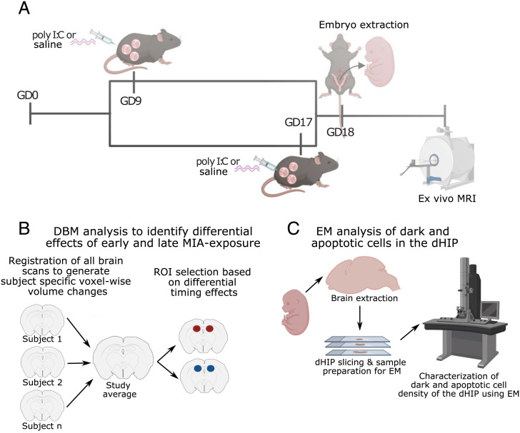
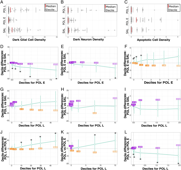
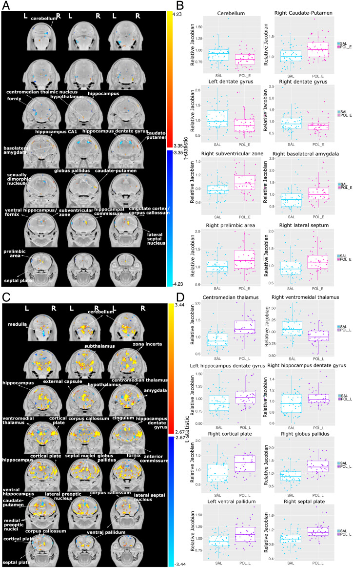
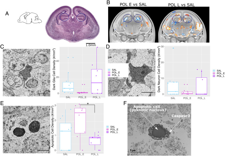
Exposure to maternal immune activation (MIA) in utero is a risk factor for neurodevelopmental and psychiatric disorders. MIA-induced deficits in adolescent and adult offspring have been well characterized; however, less is known about the effects of MIA exposure on embryo development. To address this gap, we performed high-resolution ex vivo MRI to investigate the effects of early (gestational day [GD]9) and late (GD17) MIA exposure on embryo (GD18) brain structure. We identify striking neuroanatomical changes in the embryo brain, particularly in the late-exposed offspring. We further examined the putative neuroanatomical underpinnings of MIA timing in the hippocampus using electron microscopy and identified differential effects due to MIA timing. An increase in apoptotic cell density was observed in the GD9-exposed offspring, while an increase in the density of neurons and glia with ultrastructural features reflective of increased neuroinflammation and oxidative stress was observed in GD17-exposed offspring, particularly in females. Overall, our findings integrate imaging techniques across different scales to identify differential impact of MIA timing on the earliest stages of neurodevelopment.
子宫内母体免疫激活 (MIA) 暴露是神经发育和精神疾病的风险因素。已经很好地描述了 MIA 诱导的青少年和成年后代的缺陷;然而,对于 MIA 暴露对胚胎发育的影响知之甚少。为了解决这一差距,我们进行了高分辨率的离体 MRI 研究,以调查早期(妊娠第 9 天 [GD])和晚期(GD17)MIA 暴露对胚胎(GD18)大脑结构的影响。我们发现胚胎大脑存在明显的神经解剖学变化,特别是在晚期暴露的后代中。我们进一步使用电子显微镜检查了海马体中 MIA 时间的潜在神经解剖学基础,并发现了由于 MIA 时间不同而产生的差异影响。在 GD9 暴露的后代中观察到凋亡细胞密度增加,而在 GD17 暴露的后代中观察到神经元和神经胶质密度增加,具有反映神经炎症和氧化应激增加的超微结构特征,特别是在雌性中。总的来说,我们的研究结果整合了不同尺度的成像技术,以确定 MIA 时间对神经发育最早阶段的不同影响。