Moog Nora K, Nolvi Saara, Kleih Theresa S, Styner Martin, Gilmore John H, Rasmussen Jerod M, Heim Christine M, Entringer Sonja, Wadhwa Pathik D, Buss Claudia
Charité - Universitätsmedizin Berlin, corporate member of Freie Universität Berlin and Humboldt-Universität zu Berlin, Department of Medical Psychology, Augustenburger Platz 1, 13353, Berlin, Germany.
Turku Institute for Advanced Studies, Department of Psychology and Speech-Language Pathology, University of Turku, Finland.
Neurobiol Stress. 2021 Jul 16;15:100368. doi: 10.1016/j.ynstr.2021.100368. eCollection 2021 Nov.
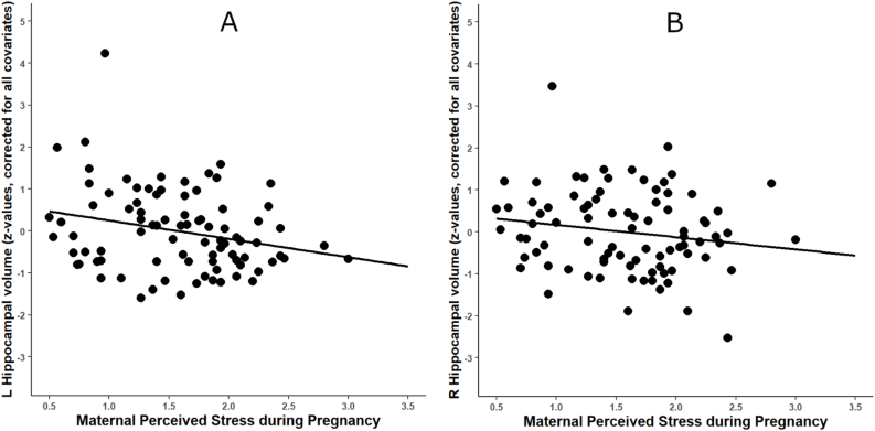
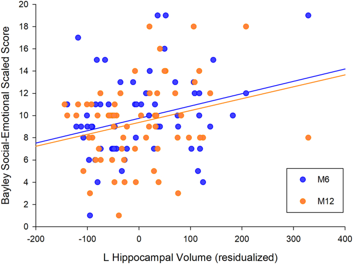
Maternal psychosocial stress during pregnancy can impact the developing fetal brain and influence offspring mental health. In this context, animal studies have identified the hippocampus and amygdala as key brain regions of interest, however, evidence in humans is sparse. We, therefore, examined the associations between maternal prenatal psychosocial stress, newborn hippocampal and amygdala volumes, and child social-emotional development. In a sample of 86 mother-child dyads, maternal perceived stress was assessed serially in early, mid and late pregnancy. Following birth, newborn (aged 5-64 postnatal days, mean: 25.8 ± 12.9) hippocampal and amygdala volume was assessed using structural magnetic resonance imaging. Infant social-emotional developmental milestones were assessed at 6- and 12-months age using the Bayley-III. After adjusting for covariates, maternal perceived stress during pregnancy was inversely associated with newborn left hippocampal volume ( = -0.26, p = .019), but not with right hippocampal ( = -0.170, = .121) or bilateral amygdala volumes (s > .5). Furthermore, newborn left hippocampal volume was positively associated with infant social-emotional development across the first year of postnatal life (B = 0.01, p = .011). Maternal perceived stress was indirectly associated with infant social-emotional development via newborn left hippocampal volume (B = -0.34, 95% CI [-0.97, -0.01]), suggesting mediation. This study provides prospective evidence in humans linking maternal psychosocial stress in pregnancy with newborn hippocampal volume and subsequent infant social-emotional development across the first year of life. These findings highlight the importance of maternal psychosocial state during pregnancy as a target amenable to interventions to prevent or attenuate its potentially unfavorable neural and behavioral consequences in the offspring.
孕期母亲的心理社会压力会影响发育中的胎儿大脑,并影响后代的心理健康。在此背景下,动物研究已确定海马体和杏仁核是关键的感兴趣脑区,然而,人类方面的证据却很少。因此,我们研究了母亲产前心理社会压力、新生儿海马体和杏仁核体积与儿童社会情感发展之间的关联。在一个包含86对母婴的样本中,在孕早期、中期和晚期连续评估母亲感知到的压力。出生后,使用结构磁共振成像评估新生儿(出生后5 - 64天,平均:25.8 ± 12.9)海马体和杏仁核的体积。在6个月和12个月大时,使用贝利婴幼儿发展量表第三版评估婴儿的社会情感发育里程碑。在调整协变量后,孕期母亲感知到的压力与新生儿左侧海马体体积呈负相关(β = -0.26,p = 0.019),但与右侧海马体(β = -0.170,p = 0.121)或双侧杏仁核体积无关(p > 0.5)。此外,新生儿左侧海马体体积与出生后第一年婴儿的社会情感发展呈正相关(B = 0.01,p = 0.011)。母亲感知到的压力通过新生儿左侧海马体体积与婴儿社会情感发展间接相关(B = -0.34,95%置信区间[-0.97, -0.01]),表明存在中介作用。这项研究为人类提供了前瞻性证据,将孕期母亲的心理社会压力与新生儿海马体体积以及出生后第一年婴儿随后的社会情感发展联系起来。这些发现凸显了孕期母亲心理社会状态作为一个可干预目标的重要性,以预防或减轻其对后代潜在的不利神经和行为后果。