• 文献检索
  • 文档翻译
  • 深度研究
  • 学术资讯
  • Suppr Zotero 插件Zotero 插件
  • 邀请有礼
  • 套餐&价格
  • 历史记录
应用&插件
Suppr Zotero 插件Zotero 插件浏览器插件Mac 客户端Windows 客户端微信小程序
定价
高级版会员购买积分包购买API积分包
服务
文献检索文档翻译深度研究API 文档MCP 服务
关于我们
关于 Suppr公司介绍联系我们用户协议隐私条款
关注我们

Suppr 超能文献

核心技术专利:CN118964589B侵权必究
粤ICP备2023148730 号-1Suppr @ 2026

文献检索

告别复杂PubMed语法,用中文像聊天一样搜索,搜遍4000万医学文献。AI智能推荐,让科研检索更轻松。

立即免费搜索

文件翻译

保留排版,准确专业,支持PDF/Word/PPT等文件格式,支持 12+语言互译。

免费翻译文档

深度研究

AI帮你快速写综述,25分钟生成高质量综述,智能提取关键信息,辅助科研写作。

立即免费体验

血小板释放的细胞外囊泡:凝血酶激活的影响。

Platelet-released extracellular vesicles: the effects of thrombin activation.

机构信息

Cardiovascular Program ICCC, Research Institute Hospital Santa Creu i Sant Pau, IIB Sant Pau, c/Sant Antoni Mª Claret 167, 08025, Barcelona, Spain.

CIBERCV Instituto de Salud Carlos III, Madrid, Spain.

出版信息

Cell Mol Life Sci. 2022 Mar 14;79(3):190. doi: 10.1007/s00018-022-04222-4.

DOI:10.1007/s00018-022-04222-4
PMID:35288766
原文链接:https://pmc.ncbi.nlm.nih.gov/articles/PMC8920058/
Abstract

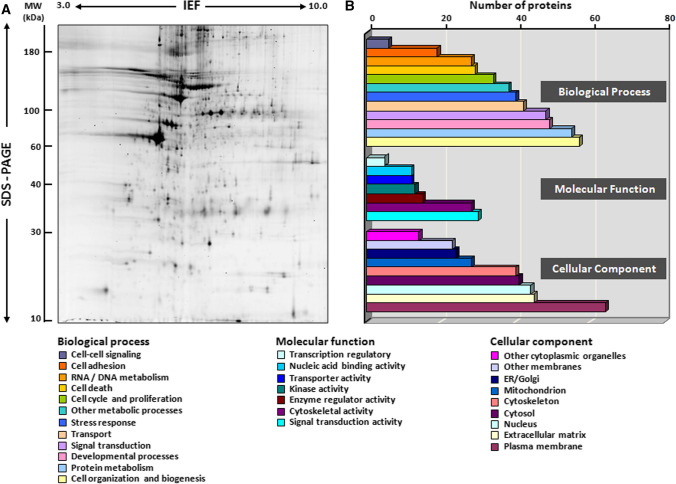
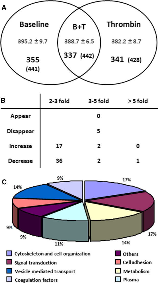
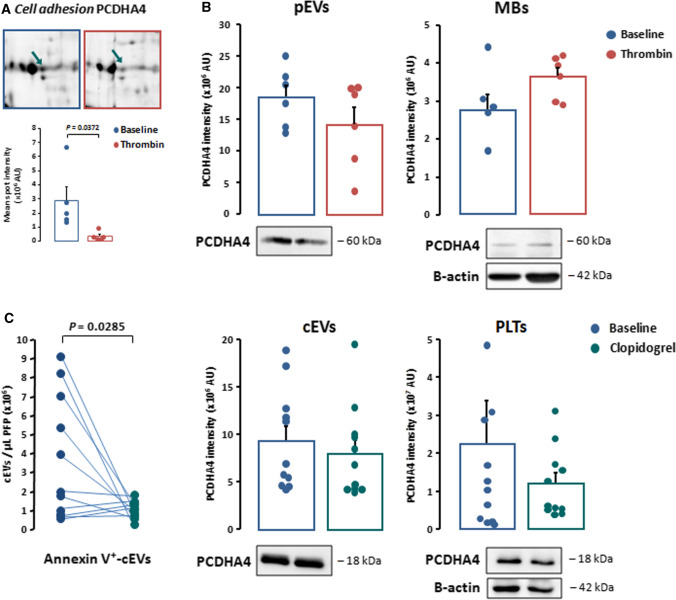
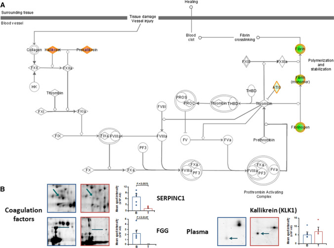
Platelets exert fundamental roles in thrombosis, inflammation, and angiogenesis, contributing to different pathologies from cardiovascular diseases to cancer. We previously reported that platelets release extracellular vesicles (pEVs) which contribute to thrombus formation. However, pEV composition remains poorly defined. Indeed, pEV quality and type, rather than quantity, may be relevant in intravascular cross-talk with either circulating or vascular cells. We aimed to define the phenotypic characteristics of pEVs released spontaneously and those induced by thrombin activation to better understand their role in disease dissemination. pEVs obtained from washed platelets from healthy donor blood were characterized by flow cytometry. pEVs from thrombin-activated platelets (T-pEVs) showed higher levels of P-selectin and active form of glycoprotein IIb/IIIa than baseline non-activated platelets (B-pEVs). Following mass spectrometry-based differential proteomic analysis, significant changes in the abundance of proteins secreted in T-pEVs compared to B-pEVs were found. These differential proteins were involved in coagulation, adhesion, cytoskeleton, signal transduction, metabolism, and vesicle-mediated transport. Interestingly, release of proteins relevant for cell adhesion, intrinsic pathway coagulation, and platelet activation signalling was significantly modified by thrombin stimulation. A novel pEV-associated protein (protocadherin-α4) was found to be significantly reduced in T-pEVs showing a shift towards increased expression in the membranes of activated platelets. In summary, platelet activation induced by thrombin triggers the shedding of pEVs with a complex proteomic pattern rich in procoagulant and proadhesive proteins. Crosstalk with other vascular and blood cells in a paracrine regulatory mode could extend the prothrombotic signalling as well as promote proteostasic changes in other cellular types.

摘要

血小板在血栓形成、炎症和血管生成中发挥着基本作用,导致从心血管疾病到癌症等不同的病理变化。我们之前报道过,血小板释放细胞外囊泡(pEVs),这有助于血栓形成。然而,pEV 的组成仍然定义不明确。事实上,pEV 的质量和类型,而不是数量,可能与循环或血管细胞的血管内相互作用有关。我们旨在确定自发性释放的 pEVs 和被凝血酶激活诱导的 pEVs 的表型特征,以更好地了解它们在疾病传播中的作用。通过流式细胞术对来自健康供体血液的血小板的 pEVs 进行了表征。与基础非激活血小板(B-pEVs)相比,来自凝血酶激活血小板(T-pEVs)的 pEVs 显示出更高水平的 P-选择素和糖蛋白 IIb/IIIa 的活性形式。在基于质谱的差异蛋白质组学分析后,发现 T-pEVs 中分泌蛋白的丰度与 B-pEVs 相比有显著变化。这些差异蛋白参与了凝血、粘附、细胞骨架、信号转导、代谢和囊泡介导的运输。有趣的是,与细胞粘附、内在途径凝血和血小板激活信号转导相关的蛋白质的释放在凝血酶刺激下显著改变。发现一种新的 pEV 相关蛋白(原钙粘蛋白-α4)在 T-pEVs 中显著减少,而在激活血小板的膜中表达增加。总之,凝血酶诱导的血小板激活触发了具有丰富促凝和促黏附蛋白的 pEVs 的脱落,其蛋白质组模式复杂。旁分泌调节模式与其他血管和血细胞的相互作用可以扩展促血栓信号,并促进其他细胞类型的蛋白稳态变化。

https://cdn.ncbi.nlm.nih.gov/pmc/blobs/0b9d/11073173/f6a9b6d174b4/18_2022_4222_Fig7_HTML.jpg
https://cdn.ncbi.nlm.nih.gov/pmc/blobs/0b9d/11073173/469e5428d2c9/18_2022_4222_Fig1_HTML.jpg
https://cdn.ncbi.nlm.nih.gov/pmc/blobs/0b9d/11073173/3935a3807b11/18_2022_4222_Fig2_HTML.jpg
https://cdn.ncbi.nlm.nih.gov/pmc/blobs/0b9d/11073173/0a0dc7a08fc1/18_2022_4222_Fig3_HTML.jpg
https://cdn.ncbi.nlm.nih.gov/pmc/blobs/0b9d/11073173/c74b2384a60c/18_2022_4222_Fig4_HTML.jpg
https://cdn.ncbi.nlm.nih.gov/pmc/blobs/0b9d/11073173/dedba31a8505/18_2022_4222_Fig5_HTML.jpg
https://cdn.ncbi.nlm.nih.gov/pmc/blobs/0b9d/11073173/88416c8c00d4/18_2022_4222_Fig6_HTML.jpg
https://cdn.ncbi.nlm.nih.gov/pmc/blobs/0b9d/11073173/f6a9b6d174b4/18_2022_4222_Fig7_HTML.jpg

相似文献

1
Platelet-released extracellular vesicles: the effects of thrombin activation.血小板释放的细胞外囊泡:凝血酶激活的影响。
Cell Mol Life Sci. 2022 Mar 14;79(3):190. doi: 10.1007/s00018-022-04222-4.
2
Platelet-derived extracellular vesicles released after trauma promote hemostasis and contribute to DVT in mice.创伤后释放的血小板衍生细胞外囊泡促进止血,并有助于小鼠深静脉血栓形成。
J Thromb Haemost. 2019 Oct;17(10):1733-1745. doi: 10.1111/jth.14563. Epub 2019 Jul 28.
3
Platelet-derived- Extracellular Vesicles Promote Hemostasis and Prevent the Development of Hemorrhagic Shock.血小板衍生的细胞外囊泡促进止血并预防出血性休克的发生。
Sci Rep. 2019 Nov 27;9(1):17676. doi: 10.1038/s41598-019-53724-y.
4
Dissecting the biochemical architecture and morphological release pathways of the human platelet extracellular vesiculome.解析人类血小板细胞外囊泡组的生化结构和形态释放途径。
Cell Mol Life Sci. 2018 Oct;75(20):3781-3801. doi: 10.1007/s00018-018-2771-6. Epub 2018 Feb 9.
5
Differential effects of physiological agonists on the proteome of platelet-derived extracellular vesicles.生理激动剂对血小板衍生细胞外囊泡蛋白质组的差异影响。
Proteomics. 2024 Aug;24(16):e2400090. doi: 10.1002/pmic.202400090.
6
The Impact of Platelet Isolation Protocol on the Release of Extracellular Vesicles.血小板分离方案对细胞外囊泡释放的影响。
Front Biosci (Landmark Ed). 2022 May 18;27(5):161. doi: 10.31083/j.fbl2705161.
7
OxLDL sensitizes platelets for increased formation of extracellular vesicles capable of finetuning macrophage gene expression.氧化低密度脂蛋白(OxLDL)使血小板变得敏感,从而增加能够微调巨噬细胞基因表达的细胞外囊泡的形成。
Eur J Cell Biol. 2023 Jun;102(2):151311. doi: 10.1016/j.ejcb.2023.151311. Epub 2023 Mar 15.
8
Differential effects of physiological agonists on the proteome of platelet-derived extracellular vesicles.生理激动剂对血小板衍生细胞外囊泡蛋白质组的差异影响。
Proteomics. 2024 Jun;24(11):e2300391. doi: 10.1002/pmic.202300391. Epub 2024 Mar 31.
9
P2Y12 antagonist ticagrelor inhibits the release of procoagulant extracellular vesicles from activated platelets.P2Y12 拮抗剂替卡格雷抑制激活的血小板释放促凝细胞外囊泡。
Cardiol J. 2019;26(6):782-789. doi: 10.5603/CJ.a2018.0045. Epub 2018 Apr 19.
10
Beyond basic characterization and omics: Immunomodulatory roles of platelet-derived extracellular vesicles unveiled by functional testing.超越基本特征描述和组学:通过功能测试揭示血小板衍生细胞外囊泡的免疫调节作用。
J Extracell Vesicles. 2024 Oct;13(10):e12513. doi: 10.1002/jev2.12513.

引用本文的文献

1
Extracellular vesicles in atherosclerosis cardiovascular disease: emerging roles and mechanisms.动脉粥样硬化性心血管疾病中的细胞外囊泡:新出现的作用和机制
Front Cardiovasc Med. 2025 Jun 24;12:1611557. doi: 10.3389/fcvm.2025.1611557. eCollection 2025.
2
Dihydrogeodin from Modulates Platelet Aggregation via Downregulation of Calcium Signaling, αIIbβ Integrins, MAPK, and PI3K/Akt Pathways.来自[具体来源未提及]的二氢地奥司明通过下调钙信号、αIIbβ整合素、丝裂原活化蛋白激酶(MAPK)和磷脂酰肌醇-3激酶/蛋白激酶B(PI3K/Akt)信号通路来调节血小板聚集。
Mar Drugs. 2025 May 17;23(5):212. doi: 10.3390/md23050212.
3
The emerging roles of platelet-derived extracellular vesicles in disease.

本文引用的文献

1
Formation of a protein corona on the surface of extracellular vesicles in blood plasma.血浆中外泌体表面形成蛋白质冠。
J Extracell Vesicles. 2021 Sep;10(11):e12140. doi: 10.1002/jev2.12140.
2
Platelet and Endothelial Activation as Potential Mechanisms Behind the Thrombotic Complications of COVID-19 Patients.血小板和内皮细胞激活作为新冠病毒肺炎患者血栓形成并发症背后的潜在机制
JACC Basic Transl Sci. 2021 Mar;6(3):202-218. doi: 10.1016/j.jacbts.2020.12.009. Epub 2021 Feb 24.
3
Platelet Proteomes, Pathways, and Phenotypes as Informants of Vascular Wellness and Disease.
血小板衍生细胞外囊泡在疾病中的新作用。
Ann Med. 2025 Dec;57(1):2499029. doi: 10.1080/07853890.2025.2499029. Epub 2025 May 3.
4
"Unraveling the Clot-Miscarriage Nexus: Mechanisms, Management, and Future Directions in Thrombosis-Related Recurrent Pregnancy Loss".解析血栓与流产的关联:血栓相关复发性流产的机制、管理及未来方向
Clin Appl Thromb Hemost. 2025 Jan-Dec;31:10760296251339421. doi: 10.1177/10760296251339421. Epub 2025 Apr 29.
5
Deciphering Platelets: Are They Cells or an Evolved Form of Extracellular Vesicles?解读血小板:它们是细胞还是细胞外囊泡的一种进化形式?
Circ Res. 2025 Feb 14;136(4):442-452. doi: 10.1161/CIRCRESAHA.124.324721. Epub 2025 Feb 13.
6
Protease-activated receptors in vascular smooth muscle cells: a bridge between thrombo-inflammation and vascular remodelling.血管平滑肌细胞中的蛋白酶激活受体:血栓炎症与血管重塑之间的桥梁。
Cell Commun Signal. 2025 Jan 31;23(1):57. doi: 10.1186/s12964-025-02066-6.
7
Toll-like Receptors 1, 3 and 7 Activate Distinct Genetic Features of NF-κB Signaling and γ-Protocadherin Expression in Human Cardiac Fibroblasts.Toll样受体1、3和7激活人心脏成纤维细胞中NF-κB信号传导和γ-原钙黏蛋白表达的不同基因特征。
Inflammation. 2025 Jan 20. doi: 10.1007/s10753-025-02238-z.
8
The protein cargo of extracellular vesicles correlates with the epigenetic aging clock of exercise sensitive DNAmFitAge.细胞外囊泡的蛋白质负载与运动敏感的DNAmFitAge表观遗传衰老时钟相关。
Biogerontology. 2025 Jan 8;26(1):35. doi: 10.1007/s10522-024-10177-9.
9
Progress Analysis of Personalized Antiplatelet Therapy in Patients with Coronary Heart Disease Undergoing Interventional Therapy.冠心病介入治疗患者个性化抗血小板治疗的进展分析
Rev Cardiovasc Med. 2024 Dec 25;25(12):462. doi: 10.31083/j.rcm2512462. eCollection 2024 Dec.
10
Delivering therapeutic RNA into the brain using extracellular vesicles.利用细胞外囊泡将治疗性RNA递送至大脑。
Mol Ther Nucleic Acids. 2024 Oct 28;35(4):102373. doi: 10.1016/j.omtn.2024.102373. eCollection 2024 Dec 10.
血小板蛋白质组学、途径和表型作为血管健康和疾病的信息源。
Arterioscler Thromb Vasc Biol. 2021 Mar;41(3):999-1011. doi: 10.1161/ATVBAHA.120.314647. Epub 2021 Jan 14.
4
Platelet Extracellular Vesicles: Beyond the Blood.血小板细胞外囊泡:超越血液的范围。
Arterioscler Thromb Vasc Biol. 2021 Jan;41(1):87-96. doi: 10.1161/ATVBAHA.120.314644. Epub 2020 Oct 8.
5
Extracellular vesicles from human plasma and serum are carriers of extravesicular cargo-Implications for biomarker discovery.人血浆和血清中的细胞外囊泡是细胞外囊泡货物的载体——对生物标志物发现的启示。
PLoS One. 2020 Aug 19;15(8):e0236439. doi: 10.1371/journal.pone.0236439. eCollection 2020.
6
Platelet-derived extracellular vesicles infiltrate and modify the bone marrow during inflammation.血小板衍生的细胞外囊泡在炎症期间浸润并改变骨髓。
Blood Adv. 2020 Jul 14;4(13):3011-3023. doi: 10.1182/bloodadvances.2020001758.
7
Liquid Biopsy of Extracellular Microvesicles Predicts Future Major Ischemic Events in Genetically Characterized Familial Hypercholesterolemia Patients.液体活检细胞外微囊泡可预测遗传性家族性高胆固醇血症患者的未来主要缺血事件。
Arterioscler Thromb Vasc Biol. 2019 Jun;39(6):1172-1181. doi: 10.1161/ATVBAHA.119.312420.
8
Minimal information for studies of extracellular vesicles 2018 (MISEV2018): a position statement of the International Society for Extracellular Vesicles and update of the MISEV2014 guidelines.细胞外囊泡研究的最低限度信息2018(MISEV2018):国际细胞外囊泡协会的立场声明及MISEV2014指南的更新
J Extracell Vesicles. 2018 Nov 23;7(1):1535750. doi: 10.1080/20013078.2018.1535750. eCollection 2018.
9
Application of Extracellular Vesicles Proteomics to Cardiovascular Disease: Guidelines, Data Analysis, and Future Perspectives.细胞外囊泡蛋白质组学在心血管疾病中的应用:指南、数据分析和未来展望。
Proteomics. 2019 Jan;19(1-2):e1800247. doi: 10.1002/pmic.201800247. Epub 2019 Jan 7.
10
Cytokine exocytosis and JAK/STAT activation in the ovary requires the vesicle trafficking regulator α-Snap.α-Snap 作为囊泡运输调节剂,在卵巢细胞因子胞吐和 JAK/STAT 激活中发挥作用。
J Cell Sci. 2018 Nov 30;131(23):jcs217638. doi: 10.1242/jcs.217638.