• 文献检索
  • 文档翻译
  • 深度研究
  • 学术资讯
  • Suppr Zotero 插件Zotero 插件
  • 邀请有礼
  • 套餐&价格
  • 历史记录
应用&插件
Suppr Zotero 插件Zotero 插件浏览器插件Mac 客户端Windows 客户端微信小程序
定价
高级版会员购买积分包购买API积分包
服务
文献检索文档翻译深度研究API 文档MCP 服务
关于我们
关于 Suppr公司介绍联系我们用户协议隐私条款
关注我们

Suppr 超能文献

核心技术专利:CN118964589B侵权必究
粤ICP备2023148730 号-1Suppr @ 2026

文献检索

告别复杂PubMed语法,用中文像聊天一样搜索,搜遍4000万医学文献。AI智能推荐,让科研检索更轻松。

立即免费搜索

文件翻译

保留排版,准确专业,支持PDF/Word/PPT等文件格式,支持 12+语言互译。

免费翻译文档

深度研究

AI帮你快速写综述,25分钟生成高质量综述,智能提取关键信息,辅助科研写作。

立即免费体验

预测新冠病毒疾病严重程度的免疫表型。

Immune phenotypes that predict COVID-19 severity.

作者信息

Liechti Thomas, Iftikhar Yaser, Mangino Massimo, Beddall Margaret, Goss Charles W, O'Halloran Jane A, Mudd Philip, Roederer Mario

机构信息

ImmunoTechnology Section, Vaccine Research Center, NIAID, NIH, USA, 20892.

Department of Twin Research & Genetic Epidemiology, King's College of London, London, UK.

出版信息

Res Sq. 2022 Mar 10:rs.3.rs-1378671. doi: 10.21203/rs.3.rs-1378671/v1.

DOI:10.21203/rs.3.rs-1378671/v1
PMID:35291290
原文链接:https://pmc.ncbi.nlm.nih.gov/articles/PMC8923110/
Abstract

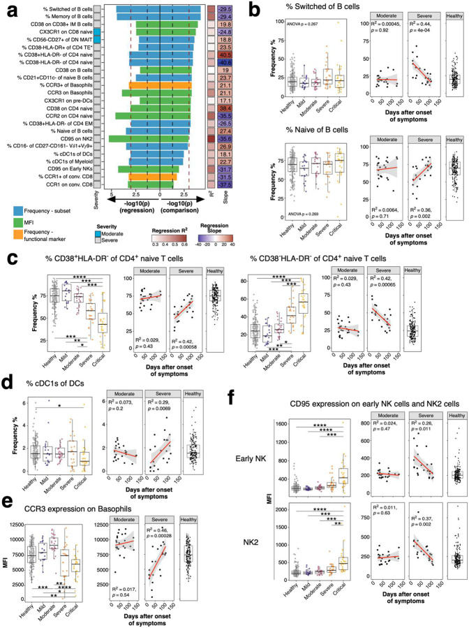
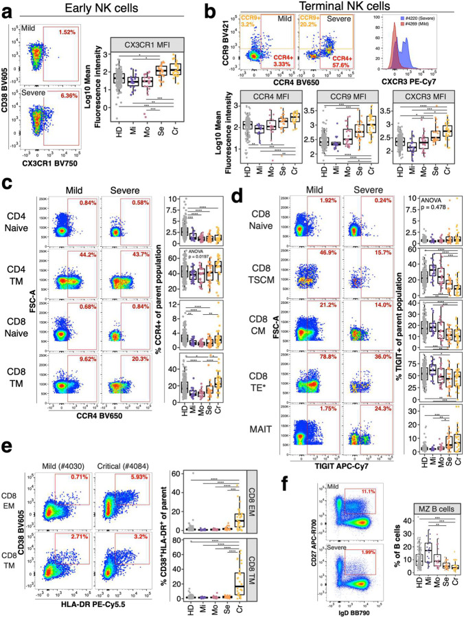
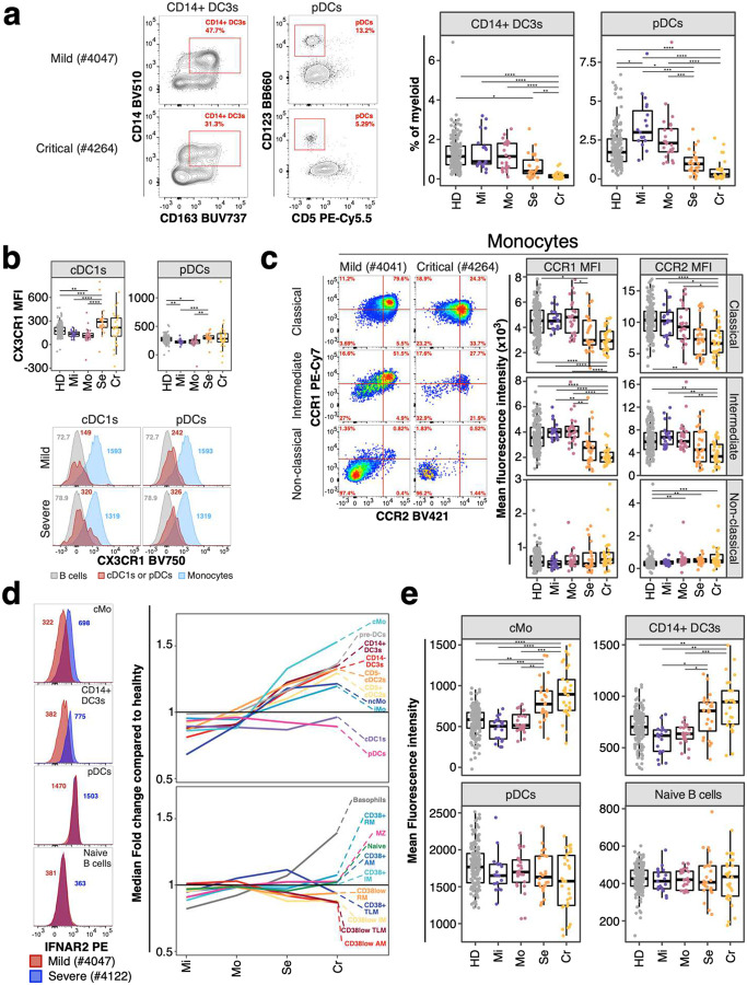
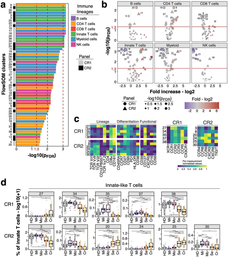
Severe COVID-19 causes profound immune perturbations, but pre-infection immune signatures contributing to severe COVID-19 remain unknown. Genome-wide association studies (GWAS) identified strong associations between severe disease and several chemokine receptors and molecules from the type I interferon pathway. Here, we define immune signatures associated with severe COVID-19 using high-dimensional flow cytometry. We measured the peripheral immune system from individuals who recovered from mild, moderate, severe or critical COVID-19 and focused only on those immune signatures returning to steady-state. Individuals that suffered from severe COVID-19 showed reduced frequencies of T cell, MAIT cell and dendritic cell (DCs) subsets and altered chemokine receptor expression on several subsets, such as reduced levels of CCR1 and CCR2 on monocyte subsets. Furthermore, we found reduced frequencies of type I interferon-producing plasmacytoid DCs and altered IFNAR2 expression on several myeloid cells in individuals recovered from severe COVID-19. Thus, these data identify potential immune mechanisms contributing to severe COVID-19.

摘要

重症新型冠状病毒肺炎(COVID-19)会引发严重的免疫紊乱,但导致重症COVID-19的感染前免疫特征仍不清楚。全基因组关联研究(GWAS)确定了重症疾病与几种趋化因子受体以及I型干扰素途径中的分子之间存在密切关联。在此,我们使用高维流式细胞术定义了与重症COVID-19相关的免疫特征。我们测量了从轻度、中度、重度或危重症COVID-19中康复的个体的外周免疫系统,并且只关注那些恢复到稳态的免疫特征。患有重症COVID-19的个体表现出T细胞、黏膜相关恒定T细胞(MAIT细胞)和树突状细胞(DCs)亚群的频率降低,并且几个亚群上的趋化因子受体表达发生改变,例如单核细胞亚群上CCR1和CCR2水平降低。此外,我们发现从重症COVID-19中康复的个体中,产生I型干扰素的浆细胞样DCs频率降低,并且几种髓样细胞上的IFNAR2表达发生改变。因此,这些数据确定了导致重症COVID-19的潜在免疫机制。

https://cdn.ncbi.nlm.nih.gov/pmc/blobs/691f/8923110/19dccf925af8/nihpp-rs1378671v1-f0006.jpg
https://cdn.ncbi.nlm.nih.gov/pmc/blobs/691f/8923110/83ccb095d3e9/nihpp-rs1378671v1-f0007.jpg
https://cdn.ncbi.nlm.nih.gov/pmc/blobs/691f/8923110/b16f2878b7d7/nihpp-rs1378671v1-f0008.jpg
https://cdn.ncbi.nlm.nih.gov/pmc/blobs/691f/8923110/05baffe61882/nihpp-rs1378671v1-f0009.jpg
https://cdn.ncbi.nlm.nih.gov/pmc/blobs/691f/8923110/ca1c8186680b/nihpp-rs1378671v1-f0010.jpg
https://cdn.ncbi.nlm.nih.gov/pmc/blobs/691f/8923110/3a8289ddf86c/nihpp-rs1378671v1-f0011.jpg
https://cdn.ncbi.nlm.nih.gov/pmc/blobs/691f/8923110/47e7c681172d/nihpp-rs1378671v1-f0012.jpg
https://cdn.ncbi.nlm.nih.gov/pmc/blobs/691f/8923110/ef90bd1df0c3/nihpp-rs1378671v1-f0013.jpg
https://cdn.ncbi.nlm.nih.gov/pmc/blobs/691f/8923110/2ac6bccd7416/nihpp-rs1378671v1-f0001.jpg
https://cdn.ncbi.nlm.nih.gov/pmc/blobs/691f/8923110/a230cb305e34/nihpp-rs1378671v1-f0002.jpg
https://cdn.ncbi.nlm.nih.gov/pmc/blobs/691f/8923110/fc88c1afc1a4/nihpp-rs1378671v1-f0003.jpg
https://cdn.ncbi.nlm.nih.gov/pmc/blobs/691f/8923110/26ebad74de51/nihpp-rs1378671v1-f0004.jpg
https://cdn.ncbi.nlm.nih.gov/pmc/blobs/691f/8923110/c1ba86975768/nihpp-rs1378671v1-f0005.jpg
https://cdn.ncbi.nlm.nih.gov/pmc/blobs/691f/8923110/19dccf925af8/nihpp-rs1378671v1-f0006.jpg

相似文献

1
Immune phenotypes that predict COVID-19 severity.预测新冠病毒疾病严重程度的免疫表型。
Res Sq. 2022 Mar 10:rs.3.rs-1378671. doi: 10.21203/rs.3.rs-1378671/v1.
2
Immune phenotypes that are associated with subsequent COVID-19 severity inferred from post-recovery samples.从康复后样本推断出与随后 COVID-19 严重程度相关的免疫表型。
Nat Commun. 2022 Nov 25;13(1):7255. doi: 10.1038/s41467-022-34638-2.
3
Integrating single-cell sequencing data with GWAS summary statistics reveals CD16+monocytes and memory CD8+T cells involved in severe COVID-19.单细胞测序数据与 GWAS 汇总统计数据的整合揭示了 CD16+单核细胞和记忆 CD8+T 细胞参与严重 COVID-19。
Genome Med. 2022 Feb 17;14(1):16. doi: 10.1186/s13073-022-01021-1.
4
Comprehensive immune cell profiling depicts an early immune response associated with severe coronavirus disease 2019 in cancer patients.全面的免疫细胞分析描绘了癌症患者与严重 2019 冠状病毒病相关的早期免疫反应。
Immunol Cell Biol. 2022 Jan;100(1):61-73. doi: 10.1111/imcb.12504. Epub 2021 Oct 27.
5
Immunological and inflammatory profiles during acute and convalescent phases of severe/ critically ill COVID-19 patients.严重/危重症 COVID-19 患者急性和康复期的免疫和炎症特征。
Int Immunopharmacol. 2021 Aug;97:107685. doi: 10.1016/j.intimp.2021.107685. Epub 2021 Apr 17.
6
Single-Cell Transcriptomic Profiling of MAIT Cells in Patients With COVID-19.单细胞转录组分析 COVID-19 患者中的 MAIT 细胞。
Front Immunol. 2021 Jul 30;12:700152. doi: 10.3389/fimmu.2021.700152. eCollection 2021.
7
Glomerular accumulation of plasmacytoid dendritic cells in active lupus nephritis: role of interleukin-18.浆细胞样树突状细胞在活动性狼疮性肾炎中的肾小球积聚:白细胞介素-18的作用
Arthritis Rheum. 2008 Jan;58(1):251-62. doi: 10.1002/art.23186.
8
Blood dendritic cells in systemic lupus erythematosus exhibit altered activation state and chemokine receptor function.系统性红斑狼疮患者血液树突状细胞呈现激活状态改变和趋化因子受体功能异常。
Ann Rheum Dis. 2010 Jul;69(7):1370-7. doi: 10.1136/ard.2009.111021. Epub 2009 Oct 22.
9
Monocytes expressing activin A and CCR2 exacerbate chronic testicular inflammation by promoting immune cell infiltration.表达激活素A和CCR2的单核细胞通过促进免疫细胞浸润加剧慢性睾丸炎症。
Hum Reprod. 2024 May 22. doi: 10.1093/humrep/deae107.
10
Increased sCD163 and sCD14 Plasmatic Levels and Depletion of Peripheral Blood Pro-Inflammatory Monocytes, Myeloid and Plasmacytoid Dendritic Cells in Patients With Severe COVID-19 Pneumonia.严重 COVID-19 肺炎患者的 sCD163 和 sCD14 血浆水平升高及外周血促炎单核细胞、髓系和浆细胞样树突状细胞耗竭。
Front Immunol. 2021 Feb 26;12:627548. doi: 10.3389/fimmu.2021.627548. eCollection 2021.

本文引用的文献

1
Long-term perturbation of the peripheral immune system months after SARS-CoV-2 infection.SARS-CoV-2 感染数月后外周免疫系统的长期紊乱。
BMC Med. 2022 Jan 14;20(1):26. doi: 10.1186/s12916-021-02228-6.
2
Efficacy of interferon beta-1a plus remdesivir compared with remdesivir alone in hospitalised adults with COVID-19: a double-bind, randomised, placebo-controlled, phase 3 trial.干扰素 β-1a 联合瑞德西韦与单用瑞德西韦治疗住院 COVID-19 成人患者的疗效比较:一项双盲、随机、安慰剂对照、3 期临床试验。
Lancet Respir Med. 2021 Dec;9(12):1365-1376. doi: 10.1016/S2213-2600(21)00384-2. Epub 2021 Oct 18.
3
The interferon landscape along the respiratory tract impacts the severity of COVID-19.
呼吸道中的干扰素景观影响着 COVID-19 的严重程度。
Cell. 2021 Sep 16;184(19):4953-4968.e16. doi: 10.1016/j.cell.2021.08.016. Epub 2021 Aug 19.
4
An updated guide for the perplexed: cytometry in the high-dimensional era.《困惑者最新指南:高维时代的细胞计数法》
Nat Immunol. 2021 Oct;22(10):1190-1197. doi: 10.1038/s41590-021-01006-z.
5
Mapping the human genetic architecture of COVID-19.绘制人类 COVID-19 遗传结构图谱。
Nature. 2021 Dec;600(7889):472-477. doi: 10.1038/s41586-021-03767-x. Epub 2021 Jul 8.
6
Longitudinal analysis reveals that delayed bystander CD8+ T cell activation and early immune pathology distinguish severe COVID-19 from mild disease.纵向分析显示,迟发性旁观者 CD8+T 细胞激活和早期免疫病理学将重症 COVID-19 与轻症疾病区分开来。
Immunity. 2021 Jun 8;54(6):1257-1275.e8. doi: 10.1016/j.immuni.2021.05.010. Epub 2021 May 16.
7
COVID-19 and the human innate immune system.新型冠状病毒肺炎与人类固有免疫系统。
Cell. 2021 Apr 1;184(7):1671-1692. doi: 10.1016/j.cell.2021.02.029. Epub 2021 Feb 16.
8
Monocytes, macrophages, dendritic cells and neutrophils: an update on lifespan kinetics in health and disease.单核细胞、巨噬细胞、树突状细胞和中性粒细胞:健康和疾病中寿命动力学的最新研究进展。
Immunology. 2021 Jul;163(3):250-261. doi: 10.1111/imm.13320. Epub 2021 Mar 15.
9
Association of Social and Demographic Factors With COVID-19 Incidence and Death Rates in the US.社会人口因素与美国 COVID-19 发病率和死亡率的关联。
JAMA Netw Open. 2021 Jan 4;4(1):e2036462. doi: 10.1001/jamanetworkopen.2020.36462.
10
Discriminating mild from critical COVID-19 by innate and adaptive immune single-cell profiling of bronchoalveolar lavages.通过对支气管肺泡灌洗液的固有和适应性免疫单细胞分析来区分轻症和重症 COVID-19。
Cell Res. 2021 Mar;31(3):272-290. doi: 10.1038/s41422-020-00455-9. Epub 2021 Jan 21.