• 文献检索
  • 文档翻译
  • 深度研究
  • 学术资讯
  • Suppr Zotero 插件Zotero 插件
  • 邀请有礼
  • 套餐&价格
  • 历史记录
应用&插件
Suppr Zotero 插件Zotero 插件浏览器插件Mac 客户端Windows 客户端微信小程序
定价
高级版会员购买积分包购买API积分包
服务
文献检索文档翻译深度研究API 文档MCP 服务
关于我们
关于 Suppr公司介绍联系我们用户协议隐私条款
关注我们

Suppr 超能文献

核心技术专利:CN118964589B侵权必究
粤ICP备2023148730 号-1Suppr @ 2026

文献检索

告别复杂PubMed语法,用中文像聊天一样搜索,搜遍4000万医学文献。AI智能推荐,让科研检索更轻松。

立即免费搜索

文件翻译

保留排版,准确专业,支持PDF/Word/PPT等文件格式,支持 12+语言互译。

免费翻译文档

深度研究

AI帮你快速写综述,25分钟生成高质量综述,智能提取关键信息,辅助科研写作。

立即免费体验

通过对支气管肺泡灌洗液的固有和适应性免疫单细胞分析来区分轻症和重症 COVID-19。

Discriminating mild from critical COVID-19 by innate and adaptive immune single-cell profiling of bronchoalveolar lavages.

机构信息

Laboratory of Respiratory Diseases and Thoracic Surgery (BREATHE), Department of Chronic Diseases and Metabolism, KU Leuven, Leuven, Belgium.

Department of Pneumology, University Hospitals Leuven, Leuven, Belgium.

出版信息

Cell Res. 2021 Mar;31(3):272-290. doi: 10.1038/s41422-020-00455-9. Epub 2021 Jan 21.

DOI:10.1038/s41422-020-00455-9
PMID:33473155
原文链接:https://pmc.ncbi.nlm.nih.gov/articles/PMC8027624/
Abstract

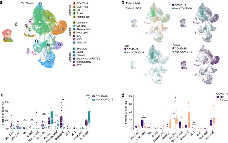
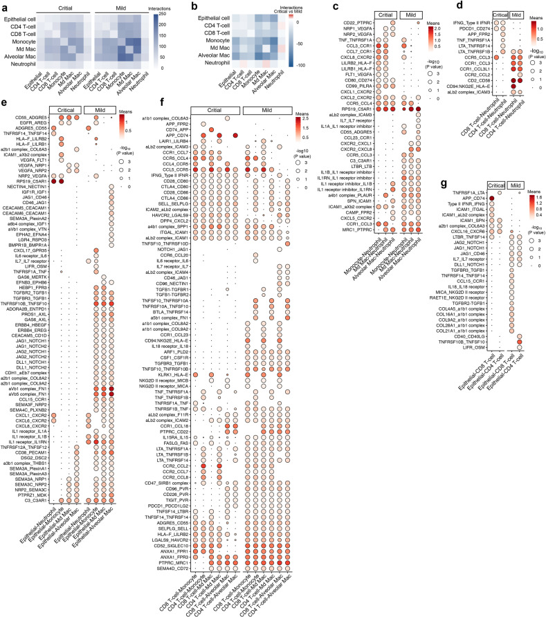
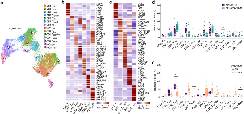
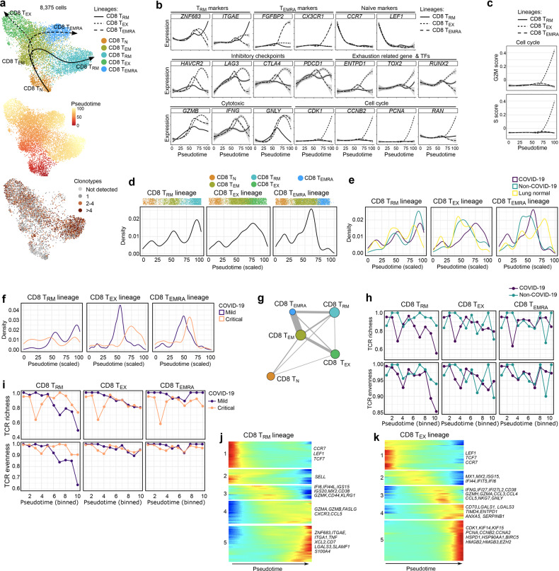
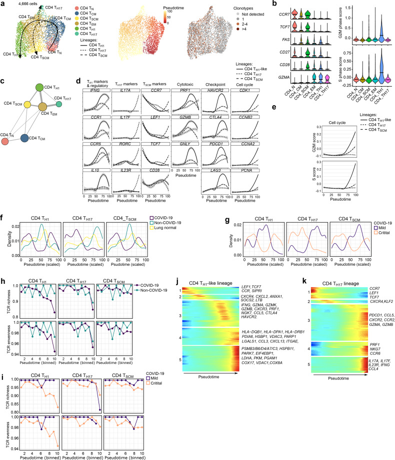
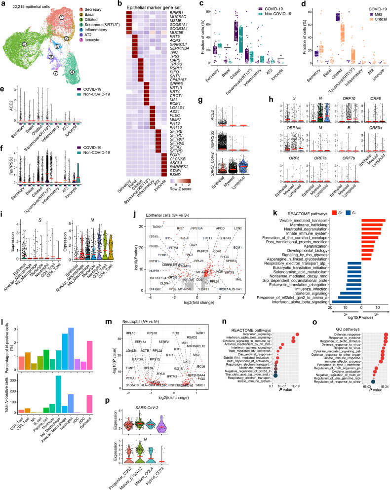
How the innate and adaptive host immune system miscommunicate to worsen COVID-19 immunopathology has not been fully elucidated. Here, we perform single-cell deep-immune profiling of bronchoalveolar lavage (BAL) samples from 5 patients with mild and 26 with critical COVID-19 in comparison to BALs from non-COVID-19 pneumonia and normal lung. We use pseudotime inference to build T-cell and monocyte-to-macrophage trajectories and model gene expression changes along them. In mild COVID-19, CD8 resident-memory (T) and CD4 T-helper-17 (T) cells undergo active (presumably antigen-driven) expansion towards the end of the trajectory, and are characterized by good effector functions, while in critical COVID-19 they remain more naïve. Vice versa, CD4 T-cells with T-helper-1 characteristics (T-like) and CD8 T-cells expressing exhaustion markers (T-like) are enriched halfway their trajectories in mild COVID-19, where they also exhibit good effector functions, while in critical COVID-19 they show evidence of inflammation-associated stress at the end of their trajectories. Monocyte-to-macrophage trajectories show that chronic hyperinflammatory monocytes are enriched in critical COVID-19, while alveolar macrophages, otherwise characterized by anti-inflammatory and antigen-presenting characteristics, are depleted. In critical COVID-19, monocytes contribute to an ATP-purinergic signaling-inflammasome footprint that could enable COVID-19 associated fibrosis and worsen disease-severity. Finally, viral RNA-tracking reveals infected lung epithelial cells, and a significant proportion of neutrophils and macrophages that are involved in viral clearance.

摘要

先天和适应性宿主免疫系统如何错误沟通导致 COVID-19 免疫病理学恶化尚未完全阐明。在这里,我们对 5 例轻度 COVID-19 和 26 例重症 COVID-19 患者的支气管肺泡灌洗液 (BAL) 样本与非 COVID-19 肺炎和正常肺的 BAL 样本进行单细胞深度免疫分析。我们使用拟时推断来构建 T 细胞和单核细胞-巨噬细胞轨迹,并对它们进行基因表达变化建模。在轻度 COVID-19 中,CD8 固有记忆 (T) 和 CD4 T 辅助 17 (T) 细胞在轨迹的末端经历积极的 (推测是抗原驱动的) 扩增,并且具有良好的效应功能,而在重症 COVID-19 中它们仍然更加幼稚。相反,在轻度 COVID-19 中,具有 T 辅助 1 特征 (T 样) 和表达衰竭标志物的 CD8 T 细胞 (T 样) 的 CD4 T 细胞在轨迹的中途富集,它们也表现出良好的效应功能,而在重症 COVID-19 中,它们在轨迹的末端表现出与炎症相关的应激迹象。单核细胞-巨噬细胞轨迹表明,慢性过度炎症性单核细胞在重症 COVID-19 中富集,而肺泡巨噬细胞,其特征是抗炎和抗原呈递特征,被耗尽。在重症 COVID-19 中,单核细胞有助于形成 ATP 嘌呤能信号-炎症小体特征,这可能导致 COVID-19 相关纤维化并使疾病严重程度恶化。最后,病毒 RNA 追踪揭示了受感染的肺上皮细胞以及参与病毒清除的大量中性粒细胞和巨噬细胞。

https://cdn.ncbi.nlm.nih.gov/pmc/blobs/b913/8027624/29cfcd54d7be/41422_2020_455_Fig8_HTML.jpg
https://cdn.ncbi.nlm.nih.gov/pmc/blobs/b913/8027624/a00f3e20a536/41422_2020_455_Fig1_HTML.jpg
https://cdn.ncbi.nlm.nih.gov/pmc/blobs/b913/8027624/e2d8c4779dc9/41422_2020_455_Fig2_HTML.jpg
https://cdn.ncbi.nlm.nih.gov/pmc/blobs/b913/8027624/34f1b8c890de/41422_2020_455_Fig3_HTML.jpg
https://cdn.ncbi.nlm.nih.gov/pmc/blobs/b913/8027624/63c936cfe8e2/41422_2020_455_Fig4_HTML.jpg
https://cdn.ncbi.nlm.nih.gov/pmc/blobs/b913/8027624/107ab5900c4b/41422_2020_455_Fig5_HTML.jpg
https://cdn.ncbi.nlm.nih.gov/pmc/blobs/b913/8027624/02c30684e82a/41422_2020_455_Fig6_HTML.jpg
https://cdn.ncbi.nlm.nih.gov/pmc/blobs/b913/8027624/07df5c314e56/41422_2020_455_Fig7_HTML.jpg
https://cdn.ncbi.nlm.nih.gov/pmc/blobs/b913/8027624/29cfcd54d7be/41422_2020_455_Fig8_HTML.jpg

相似文献

1
Discriminating mild from critical COVID-19 by innate and adaptive immune single-cell profiling of bronchoalveolar lavages.通过对支气管肺泡灌洗液的固有和适应性免疫单细胞分析来区分轻症和重症 COVID-19。
Cell Res. 2021 Mar;31(3):272-290. doi: 10.1038/s41422-020-00455-9. Epub 2021 Jan 21.
2
Single-cell multi-omics reveals dyssynchrony of the innate and adaptive immune system in progressive COVID-19.单细胞多组学揭示了进展期新冠肺炎中固有免疫系统和适应性免疫系统的不同步性。
Nat Commun. 2022 Jan 21;13(1):440. doi: 10.1038/s41467-021-27716-4.
3
Single-Cell Transcriptome Analysis Highlights a Role for Neutrophils and Inflammatory Macrophages in the Pathogenesis of Severe COVID-19.单细胞转录组分析强调中性粒细胞和炎症性巨噬细胞在严重 COVID-19 发病机制中的作用。
Cells. 2020 Oct 29;9(11):2374. doi: 10.3390/cells9112374.
4
Single-cell transcriptomics identifies pathogenic T-helper 17.1 cells and pro-inflammatory monocytes in immune checkpoint inhibitor-related pneumonitis.单细胞转录组学鉴定出免疫检查点抑制剂相关肺炎中的致病性辅助性 T 细胞 17.1 细胞和促炎单核细胞。
J Immunother Cancer. 2022 Sep;10(9). doi: 10.1136/jitc-2022-005323.
5
Phenotypic analysis of lymphocytes and monocytes/macrophages in peripheral blood and bronchoalveolar lavage fluid from patients with pulmonary sarcoidosis.结节病患者外周血及支气管肺泡灌洗液中淋巴细胞和单核细胞/巨噬细胞的表型分析。
Thorax. 1999 Apr;54(4):339-46. doi: 10.1136/thx.54.4.339.
6
Innate-like Gene Expression of Lung-Resident Memory CD8 T Cells during Experimental Human Influenza: A Clinical Study.实验性人类流感期间肺驻留记忆性CD8 T细胞的固有样基因表达:一项临床研究
Am J Respir Crit Care Med. 2021 Oct 1;204(7):826-841. doi: 10.1164/rccm.202103-0620OC.
7
Single-cell analysis reveals cell communication triggered by macrophages associated with the reduction and exhaustion of CD8 T cells in COVID-19.单细胞分析揭示了与 COVID-19 中 CD8 T 细胞减少和耗竭相关的巨噬细胞触发的细胞通讯。
Cell Commun Signal. 2021 Jul 8;19(1):73. doi: 10.1186/s12964-021-00754-7.
8
Critical COVID-19 is associated with distinct leukocyte phenotypes and transcriptome patterns.危重新冠肺炎与独特的白细胞表型和转录组模式相关。
J Intern Med. 2021 Sep;290(3):677-692. doi: 10.1111/joim.13310. Epub 2021 Jun 3.
9
Epigenetic Landscapes of Single-Cell Chromatin Accessibility and Transcriptomic Immune Profiles of T Cells in COVID-19 Patients.单细胞染色质可及性的表观遗传学景观和 COVID-19 患者 T 细胞的转录组免疫特征。
Front Immunol. 2021 Feb 24;12:625881. doi: 10.3389/fimmu.2021.625881. eCollection 2021.
10
Integrating single-cell sequencing data with GWAS summary statistics reveals CD16+monocytes and memory CD8+T cells involved in severe COVID-19.单细胞测序数据与 GWAS 汇总统计数据的整合揭示了 CD16+单核细胞和记忆 CD8+T 细胞参与严重 COVID-19。
Genome Med. 2022 Feb 17;14(1):16. doi: 10.1186/s13073-022-01021-1.

引用本文的文献

1
Immune signatures of SARS-CoV-2 infection resolution in human lung tissues.人类肺组织中新冠病毒感染消退的免疫特征
PLoS Pathog. 2025 Sep 8;21(9):e1013469. doi: 10.1371/journal.ppat.1013469. eCollection 2025 Sep.
2
Single-cell transcriptome analysis of bronchoalveolar lavage during early SARS-CoV-2 infection.新型冠状病毒2型(SARS-CoV-2)早期感染期间支气管肺泡灌洗的单细胞转录组分析
Microbiol Spectr. 2025 Sep 2;13(9):e0271524. doi: 10.1128/spectrum.02715-24. Epub 2025 Jul 31.
3
A single-cell transcriptomic atlas reveals senescence and inflammation in the post-tuberculosis human lung.

本文引用的文献

1
A molecular cell atlas of the human lung from single-cell RNA sequencing.人类肺部单细胞 RNA 测序的分子细胞图谱。
Nature. 2020 Nov;587(7835):619-625. doi: 10.1038/s41586-020-2922-4. Epub 2020 Nov 18.
2
Elevated Calprotectin and Abnormal Myeloid Cell Subsets Discriminate Severe from Mild COVID-19.血清钙卫蛋白和异常髓系细胞亚群可区分 COVID-19 重症与轻症。
Cell. 2020 Sep 17;182(6):1401-1418.e18. doi: 10.1016/j.cell.2020.08.002. Epub 2020 Aug 5.
3
Severe COVID-19 Is Marked by a Dysregulated Myeloid Cell Compartment.严重的 COVID-19 以髓系细胞失调为特征。
单细胞转录组图谱揭示了肺结核后人类肺部的衰老和炎症。
Nat Microbiol. 2025 Jul 14. doi: 10.1038/s41564-025-02050-3.
4
Multiscale dynamic immunomodulation by a nanoemulsified Trojan-TLR7/8 adjuvant for robust protection against heterologous pandemic and endemic viruses.纳米乳化特洛伊木马-TLR7/8佐剂的多尺度动态免疫调节作用,用于对异源大流行和地方性病毒的强大保护。
Cell Mol Immunol. 2025 Jun 25. doi: 10.1038/s41423-025-01306-6.
5
Aspergillus fumigatus biology, immunopathogenicity and drug resistance.烟曲霉的生物学特性、免疫致病性及耐药性。
Nat Rev Microbiol. 2025 May 2. doi: 10.1038/s41579-025-01180-z.
6
Unveiling the heterogeneity and immunotherapy potency of tumor-associated neutrophils in the tumor microenvironment of gastric cancer.揭示胃癌肿瘤微环境中肿瘤相关中性粒细胞的异质性和免疫治疗潜力。
BMC Gastroenterol. 2025 Apr 28;25(1):303. doi: 10.1186/s12876-025-03920-0.
7
SARS-CoV-2 infection results in a unique lung proteome long after virus resolution in the hamster.在仓鼠体内,SARS-CoV-2感染在病毒清除很久之后会导致独特的肺部蛋白质组。
Npj Viruses. 2024 Aug 24;2(1):40. doi: 10.1038/s44298-024-00049-x.
8
Splenic CD169Tim4 Marginal Metallophilic Macrophages Are Essential for Wound Healing After Myocardial Infarction.脾脏CD169Tim4边缘嗜金属巨噬细胞对心肌梗死后的伤口愈合至关重要。
Circulation. 2025 Jun 17;151(24):1712-1729. doi: 10.1161/CIRCULATIONAHA.124.071772. Epub 2025 Apr 28.
9
The peptidoglycan of can persist in discrete tissues and cause systemic responses consistent with chronic illness.[此处原文中“of”后缺少具体内容]的肽聚糖可在离散组织中持续存在,并引发与慢性病一致的全身反应。
Sci Transl Med. 2025 Apr 23;17(795):eadr2955. doi: 10.1126/scitranslmed.adr2955.
10
Longitudinal Changes in Peripheral and Alveolar Monocyte and Inflammatory Biomarkers are Distinct in Hypercapnia Patients Following Pulmonary Sepsis-Induced ARDS.肺脓毒症诱导的急性呼吸窘迫综合征(ARDS)后高碳酸血症患者外周血和肺泡单核细胞及炎症生物标志物的纵向变化是不同的。
J Inflamm Res. 2025 Apr 16;18:5217-5233. doi: 10.2147/JIR.S508311. eCollection 2025.
Cell. 2020 Sep 17;182(6):1419-1440.e23. doi: 10.1016/j.cell.2020.08.001. Epub 2020 Aug 5.
4
Adaptive immune responses to SARS-CoV-2 infection in severe versus mild individuals.严重与轻度个体对 SARS-CoV-2 感染的适应性免疫反应。
Signal Transduct Target Ther. 2020 Aug 14;5(1):156. doi: 10.1038/s41392-020-00263-y.
5
Single-cell landscape of immunological responses in patients with COVID-19.COVID-19 患者免疫反应的单细胞景观。
Nat Immunol. 2020 Sep;21(9):1107-1118. doi: 10.1038/s41590-020-0762-x. Epub 2020 Aug 12.
6
A Single-Cell Atlas of the Human Healthy Airways.人类健康气道单细胞图谱
Am J Respir Crit Care Med. 2020 Dec 15;202(12):1636-1645. doi: 10.1164/rccm.201911-2199OC.
7
Immunophenotyping of COVID-19 and influenza highlights the role of type I interferons in development of severe COVID-19.COVID-19 和流感的免疫表型分析突出了 I 型干扰素在 COVID-19 重症发展中的作用。
Sci Immunol. 2020 Jul 10;5(49). doi: 10.1126/sciimmunol.abd1554.
8
COVID-19 severity correlates with airway epithelium-immune cell interactions identified by single-cell analysis.单细胞分析鉴定的 COVID-19 严重程度与气道上皮-免疫细胞相互作用相关。
Nat Biotechnol. 2020 Aug;38(8):970-979. doi: 10.1038/s41587-020-0602-4. Epub 2020 Jun 26.
9
A pan-cancer blueprint of the heterogeneous tumor microenvironment revealed by single-cell profiling.单细胞分析揭示的异质性肿瘤微环境泛癌蓝图。
Cell Res. 2020 Sep;30(9):745-762. doi: 10.1038/s41422-020-0355-0. Epub 2020 Jun 19.
10
A single-cell atlas of the peripheral immune response in patients with severe COVID-19.严重 COVID-19 患者外周免疫反应的单细胞图谱。
Nat Med. 2020 Jul;26(7):1070-1076. doi: 10.1038/s41591-020-0944-y. Epub 2020 Jun 8.