• 文献检索
  • 文档翻译
  • 深度研究
  • 学术资讯
  • Suppr Zotero 插件Zotero 插件
  • 邀请有礼
  • 套餐&价格
  • 历史记录
应用&插件
Suppr Zotero 插件Zotero 插件浏览器插件Mac 客户端Windows 客户端微信小程序
定价
高级版会员购买积分包购买API积分包
服务
文献检索文档翻译深度研究API 文档MCP 服务
关于我们
关于 Suppr公司介绍联系我们用户协议隐私条款
关注我们

Suppr 超能文献

核心技术专利:CN118964589B侵权必究
粤ICP备2023148730 号-1Suppr @ 2026

文献检索

告别复杂PubMed语法,用中文像聊天一样搜索,搜遍4000万医学文献。AI智能推荐,让科研检索更轻松。

立即免费搜索

文件翻译

保留排版,准确专业,支持PDF/Word/PPT等文件格式,支持 12+语言互译。

免费翻译文档

深度研究

AI帮你快速写综述,25分钟生成高质量综述,智能提取关键信息,辅助科研写作。

立即免费体验

胎儿中枢神经系统生物测量在先天性心脏病与非先天性心脏病中的应用。

Central nervous system biometry in fetuses with and without congenital heart diseases.

机构信息

Department of Obstetrics and Gynecology, Christophorus-Kliniken GmbH, Coesfeld, Germany.

Department of Maternal-Fetal Medicine, Institute Clinic of Gynecology, Obstetrics and Neonatology (ICGON), Hospital Clinic-IDIBAPS, University of Barcelona and Centre for Biomedical Research on Rare Diseases (CIBER-ER), Barcelona, Spain.

出版信息

Arch Gynecol Obstet. 2022 Dec;306(6):1885-1890. doi: 10.1007/s00404-022-06484-6. Epub 2022 Mar 17.

DOI:10.1007/s00404-022-06484-6
PMID:35296918
原文链接:https://pmc.ncbi.nlm.nih.gov/articles/PMC9633519/
Abstract

OBJECTIVE

To compare the fetal brain structures assessed in routine sonographic scans during the second and third trimesters in fetuses with and without congenital heart disease (CHD).

METHODS

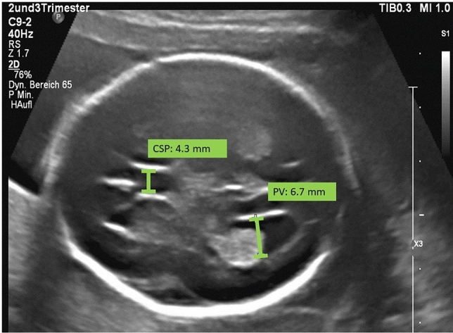
This is a retrospective cross-sectional single-center study. We measured the head circumference (HC), the transversal diameter of the cerebellum (TCD) and the sizes of the cisterna magna (CM), the cavum septi pellucidi (CSP) and the posterior ventricles (PV) between 20 and 41 weeks of gestation. We compared 160 fetuses with CHD (case group) to 160 fetuses of normal pregnancies (control group). Every patient was matched with a control, considering the gestational age at which the ultrasound was performed. We divided the CHD group into 3 subgroups: retrograde flow in the aortic arch (group 1), right heart anomaly with the antegrade flow in the aortic arch (group 2) and other CHDs with the antegrade flow in the aortic arch (group 3).

RESULTS

The mean width of the PV was larger in fetuses of groups 1 and 3 in comparison to the control group (P < 0.001, P = 0.022; respectively). We found that the APGAR score at 5 min (P < 0.001, P < 0.001; respectively) and gestational age at delivery (P = 0.006, P = 0.001; respectively) were inferior in groups 1 and 3 compared to controls.

CONCLUSIONS

Central nervous system biometry is altered in fetuses with CHD. PV is enlarged in CHD fetuses especially with decreased oxygen levels in the aortic arch.

摘要

目的

比较在胎儿第二和第三孕期常规超声扫描中评估有先天性心脏病(CHD)和无先天性心脏病胎儿的脑结构。

方法

这是一项回顾性的横断面单中心研究。我们测量了头围(HC)、小脑横径(TCD)和小脑延髓池(CM)、透明隔腔(CSP)和后颅窝(PV)的大小,在 20 至 41 孕周进行。我们将 160 例 CHD 胎儿(病例组)与 160 例正常妊娠胎儿(对照组)进行比较。每个患者都与一名对照者相匹配,考虑到进行超声检查的胎龄。我们将 CHD 组分为 3 个亚组:主动脉弓逆行血流(第 1 组)、主动脉弓前向血流右心异常(第 2 组)和其他主动脉弓前向血流 CHD(第 3 组)。

结果

与对照组相比,第 1 组和第 3 组胎儿的 PV 平均宽度更大(P<0.001,P=0.022;分别)。我们发现,第 1 组和第 3 组的 5 分钟 APGAR 评分(P<0.001,P<0.001;分别)和分娩时的胎龄(P=0.006,P=0.001;分别)均低于对照组。

结论

CHD 胎儿的中枢神经系统生物测量值发生改变。PV 在 CHD 胎儿中增大,尤其是在主动脉弓氧水平降低时。

https://cdn.ncbi.nlm.nih.gov/pmc/blobs/b188/9633519/ccf67973447e/404_2022_6484_Fig1_HTML.jpg

相似文献

1
Central nervous system biometry in fetuses with and without congenital heart diseases.胎儿中枢神经系统生物测量在先天性心脏病与非先天性心脏病中的应用。
Arch Gynecol Obstet. 2022 Dec;306(6):1885-1890. doi: 10.1007/s00404-022-06484-6. Epub 2022 Mar 17.
2
Dilated cavum septi pellucidi in fetuses with microdeletion 22q11.22q11微缺失胎儿的透明隔腔增宽
Prenat Diagn. 2016 Oct;36(10):911-915. doi: 10.1002/pd.4911. Epub 2016 Aug 30.
3
Frontal lobe growth is impaired in fetuses with congenital heart disease.先天性心脏病胎儿额叶发育受损。
Ultrasound Obstet Gynecol. 2021 May;57(5):776-782. doi: 10.1002/uog.22127. Epub 2021 Apr 13.
4
Head Biometry in Fetuses with Isolated Congenital Heart Disease.胎儿孤立性先天性心脏病的头部生物测量。
Ultraschall Med. 2020 Feb;41(1):69-76. doi: 10.1055/a-0796-6502. Epub 2018 Nov 21.
5
Head growth in fetuses with isolated congenital heart defects: lack of influence of aortic arch flow and ascending aorta oxygen saturation.孤立性先天性心脏病胎儿的头部生长:主动脉弓血流和升主动脉血氧饱和度的影响缺失
Ultrasound Obstet Gynecol. 2016 Sep;48(3):357-64. doi: 10.1002/uog.15980. Epub 2016 Aug 2.
6
The cavum septi pellucidi in euploid and aneuploid fetuses.正常胎儿和染色体异常胎儿的透明隔腔。
Ultrasound Obstet Gynecol. 2013 Aug;42(2):156-60. doi: 10.1002/uog.12393.
7
Ultrasound measurement of the fetal cavum septi pellucidi.胎儿透明隔腔的超声测量
Ultrasound Obstet Gynecol. 1998 Dec;12(6):419-21. doi: 10.1046/j.1469-0705.1998.12060419.x.
8
Sonographic measurement of the fetal cerebellum, cisterna magna, and cavum septum pellucidum in normal fetuses in the second and third trimesters of pregnancy.孕期第二和第三阶段正常胎儿小脑、枕大池及透明隔腔的超声测量
J Clin Ultrasound. 2003 May;31(4):194-200. doi: 10.1002/jcu.10163.
9
Enlarged cavum septi pellucidi Z-scores in fetuses with trisomy 18.18 三体胎儿中透明隔腔 Z 评分增大。
J Matern Fetal Neonatal Med. 2022 Mar;35(5):981-986. doi: 10.1080/14767058.2020.1742321. Epub 2020 Apr 14.
10
Longitudinal changes in fetal head biometry and fetoplacental circulation in fetuses with congenital heart defects.先天性心脏病胎儿头围生物测量和胎胎盘循环的纵向变化。
Acta Obstet Gynecol Scand. 2022 Sep;101(9):987-995. doi: 10.1111/aogs.14401. Epub 2022 Jun 20.

引用本文的文献

1
Prenatal diagnosis of cerebellar hypoplasia in fetal ultrasound using deep learning under the constraint of the anatomical structures of the cerebellum and cistern.在小脑及脑池解剖结构的约束下,利用深度学习进行胎儿超声小脑发育不全的产前诊断。
Pediatr Radiol. 2025 Sep 5. doi: 10.1007/s00247-025-06376-2.
2
Adrenal gland size in fetuses with congenital heart disease.患有先天性心脏病胎儿的肾上腺大小。
J Perinat Med. 2025 Jan 7;53(3):387-393. doi: 10.1515/jpm-2024-0402. Print 2025 Mar 26.
3
Fetal periventricular pseudocysts: is MRI evaluation needed? What is the long-term neurodevelopmental outcome? Systematic review and meta-analysis.

本文引用的文献

1
Head Biometry in Fetuses with Isolated Congenital Heart Disease.胎儿孤立性先天性心脏病的头部生物测量。
Ultraschall Med. 2020 Feb;41(1):69-76. doi: 10.1055/a-0796-6502. Epub 2018 Nov 21.
2
Fetal brain development in diabetic pregnancies and normal controls.糖尿病妊娠与正常对照中的胎儿大脑发育
J Perinat Med. 2018 Sep 25;46(7):797-803. doi: 10.1515/jpm-2017-0341.
3
Relationship Between Cavum Septi Pellucidi Measurements and Fetal Hypoplastic Left Heart Syndrome or Dextro-Transposition of the Great Arteries.透明隔腔测量值与胎儿左心发育不全综合征或大动脉右位转位之间的关系。
胎儿脑室周围假性囊肿:是否需要进行磁共振成像(MRI)评估?长期神经发育结局如何?系统评价与荟萃分析
Arch Gynecol Obstet. 2023 Jun;307(6):1697-1711. doi: 10.1007/s00404-022-06624-y. Epub 2022 Jun 8.
J Ultrasound Med. 2018 Jul;37(7):1673-1680. doi: 10.1002/jum.14515. Epub 2017 Dec 27.
4
Congenital Heart Defects and Measures of Prenatal Brain Growth: A Systematic Review.先天性心脏病与产前脑生长测量:一项系统评价
Pediatr Neurol. 2017 Jul;72:7-18.e1. doi: 10.1016/j.pediatrneurol.2017.03.014. Epub 2017 Apr 1.
5
Fetal ventriculomegaly: Diagnosis, treatment, and future directions.胎儿脑室扩大:诊断、治疗及未来方向。
Childs Nerv Syst. 2017 Jul;33(7):1113-1123. doi: 10.1007/s00381-017-3441-y. Epub 2017 May 16.
6
Mild fetal cerebral ventriculomegaly: prevalence, characteristics, and utility of ancillary testing in cases presenting to a tertiary referral center.轻度胎儿脑室扩大:三级转诊中心就诊病例的患病率、特征及辅助检查的作用
Prenat Diagn. 2017 Jul;37(7):647-657. doi: 10.1002/pd.5057. Epub 2017 Jun 1.
7
Clinical significance of slightly lateral ventricular enlargement in healthy infants.健康婴儿轻度侧脑室扩大的临床意义
J Matern Fetal Neonatal Med. 2017 Jun;30(12):1393-1396. doi: 10.1080/14767058.2016.1214704. Epub 2016 Aug 10.
8
Fetal brain imaging in isolated congenital heart defects - a systematic review and meta-analysis.孤立性先天性心脏病的胎儿脑成像——一项系统综述和荟萃分析
Prenat Diagn. 2016 Jul;36(7):601-13. doi: 10.1002/pd.4842. Epub 2016 Jun 19.
9
Mid-gestation brain Doppler and head biometry in fetuses with congenital heart disease predict abnormal brain development at birth.先天性心脏病胎儿的孕中期脑多普勒检查和头部生物测量可预测出生时脑发育异常。
Ultrasound Obstet Gynecol. 2016 Jan;47(1):65-73. doi: 10.1002/uog.14919.
10
Brain abnormalities and neurodevelopmental delay in congenital heart disease: systematic review and meta-analysis.先天性心脏病的脑异常和神经发育迟缓:系统评价和荟萃分析。
Ultrasound Obstet Gynecol. 2014 Jan;43(1):14-24. doi: 10.1002/uog.12526. Epub 2013 Dec 10.