Dept of Thoracic Diseases, G.B. Morgagni Hospital/University of Bologna, Forlì, Italy
Dept of Pathology, University Vita-Salute, Milan and San Raffaele Scientific Institute, Milan, Italy.
Eur Respir J. 2022 Oct 6;60(4). doi: 10.1183/13993003.02411-2021. Print 2022 Oct.
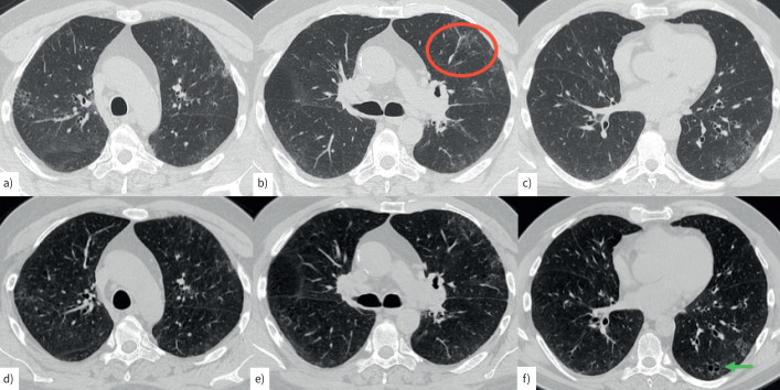
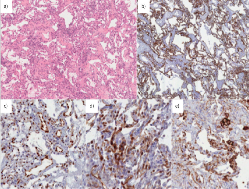
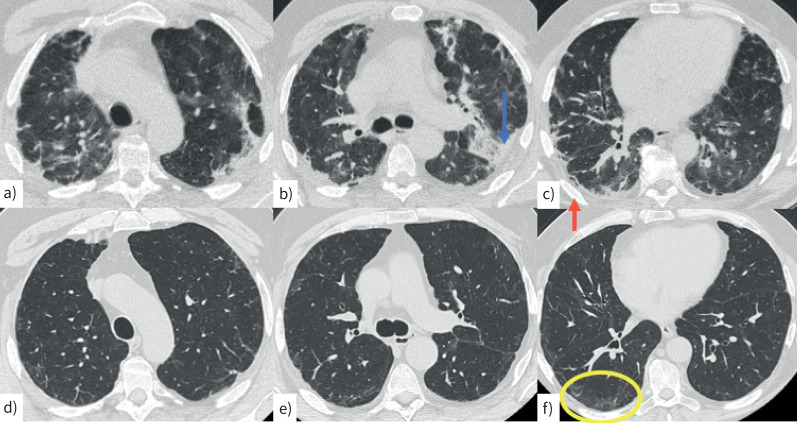
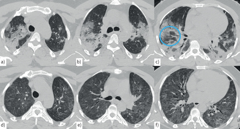
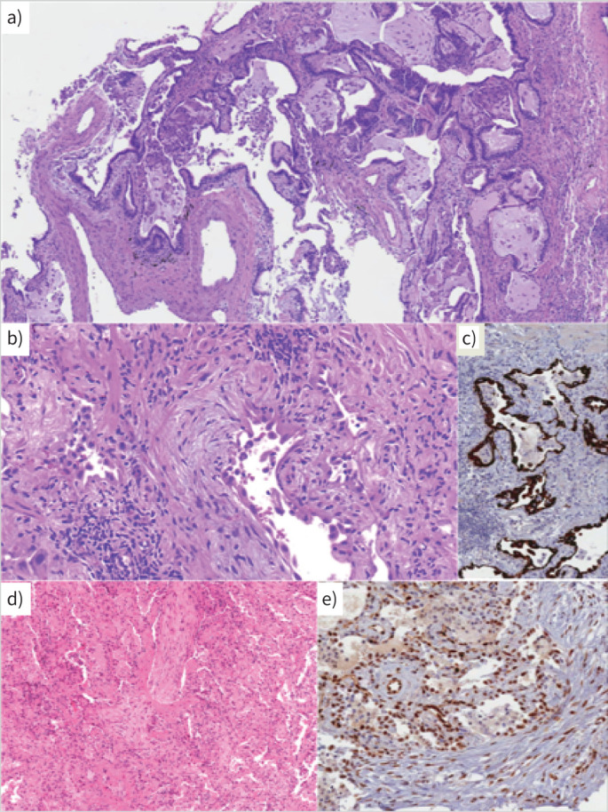
Some patients experience pulmonary sequelae after SARS-CoV-2 infection, ranging from self-limited abnormalities to major lung diseases. Morphological analysis of lung tissue may help our understanding of pathogenic mechanisms and help to provide consistent personalised management. The aim of this study was to ascertain morphological and immunomolecular features of lung tissue. Transbronchial lung cryobiopsy was carried out in patients with persistent symptoms and computed tomography suggestive of residual lung disease after recovery from SARS-CoV-2 infection. 164 patients were referred for suspected pulmonary sequelae after COVID-19; 10 patients with >5% parenchymal lung disease underwent lung biopsy. The histological pattern of lung disease was not homogeneous and three different case clusters could be identified, which was mirrored by their clinical and radiological features. Cluster 1 ("chronic fibrosing") was characterised by post-infection progression of pre-existing interstitial pneumonias. Cluster 2 ("acute/subacute injury") was characterised by different types and grades of lung injury, ranging from organising pneumonia and fibrosing nonspecific interstitial pneumonia to diffuse alveolar damage. Cluster 3 ("vascular changes") was characterised by diffuse vascular increase, dilatation and distortion (capillaries and venules) within otherwise normal parenchyma. Clusters 2 and 3 had immunophenotypical changes similar to those observed in early/mild COVID-19 pneumonias (abnormal expression of STAT3 in hyperplastic pneumocytes and PD-L1, IDO and STAT3 in endothelial cells). This is the first study correlating histological/immunohistochemical patterns with clinical and radiological pictures of patients with post-COVID lung disease. Different phenotypes with potentially different underlying pathogenic mechanisms have been identified.
一些患者在感染 SARS-CoV-2 后会出现肺部后遗症,从自限性异常到重大肺部疾病不等。对肺部组织进行形态学分析有助于我们了解发病机制,并有助于提供一致的个性化管理。本研究旨在确定肺部组织的形态学和免疫分子特征。对持续有症状且从 SARS-CoV-2 感染中康复后 CT 显示有残留肺部疾病的患者进行经支气管肺冷冻活检。有 164 名患者因疑似 COVID-19 后肺部后遗症而被转介;10 名肺部实质疾病>5%的患者接受了肺活检。肺部疾病的组织学模式并不均匀,可识别出三种不同的病例群,这与其临床和影像学特征相吻合。簇 1(“慢性纤维化”)的特征是感染后先前存在的间质性肺炎进展。簇 2(“急性/亚急性损伤”)的特征是不同类型和程度的肺损伤,从机化性肺炎和纤维化非特异性间质性肺炎到弥漫性肺泡损伤不等。簇 3(“血管变化”)的特征是弥漫性血管增加、扩张和变形(毛细血管和小静脉),而其他正常的实质不受影响。簇 2 和簇 3 具有与早期/轻度 COVID-19 肺炎观察到的免疫表型变化相似的特征(增生性肺泡细胞中 STAT3 异常表达,内皮细胞中 PD-L1、IDO 和 STAT3 异常表达)。这是第一项将组织学/免疫组织化学模式与 COVID-19 后肺部疾病患者的临床和影像学图片相关联的研究。已经确定了不同的表型,它们可能具有不同的潜在发病机制。