Nancyclotep Molecular and Experimental Imaging Platform, CHRU-Nancy, 05 rue du Morvan, 54500, Vandoeuvre-Les-Nancy, France.
Lorraine University, INSERM, IADI UMR 1254, Nancy, France.
Cancer Imaging. 2022 Mar 18;22(1):16. doi: 10.1186/s40644-022-00454-6.
This translational study explores multi-tracer PET imaging for the non-invasive detection of the IDH1 mutation which is a positive prognostic factor in glioma.
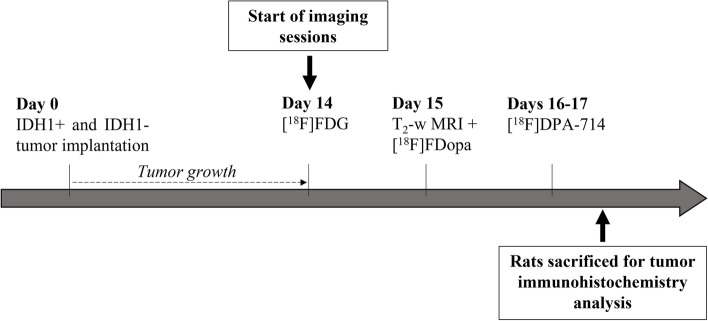
U87 human high-grade glioma (HGG) isogenic cell lines with or without the IDH1 mutation (CRISP/Cas9 method) were stereotactically grafted into rat brains, and examined, in vitro, in vivo and ex vivo. PET imaging sessions, with radiotracers specific for glycolytic metabolism ([F]FDG), amino acid metabolism ([F]FDopa), and inflammation ([F]DPA-714), were performed sequentially during 3-4 days. The in vitro radiotracer uptake was expressed as percent per million cells. For each radiotracer examined in vivo, static analyses included the maximal and mean tumor-to-background ratio (TBR and TBR) and metabolic tumor volume (MTV). Dynamic analyses included the distribution volume ratio (DVR) and the relative residence time (RRT) extracted from a reference Logan model. Ex vivo analyses consisted of immunological analyses.
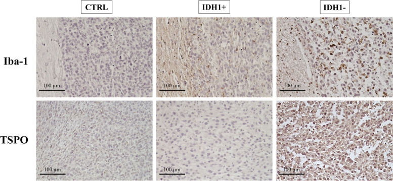
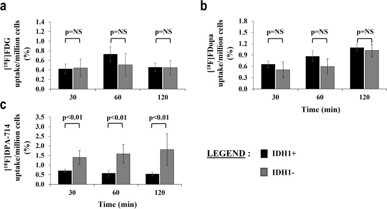
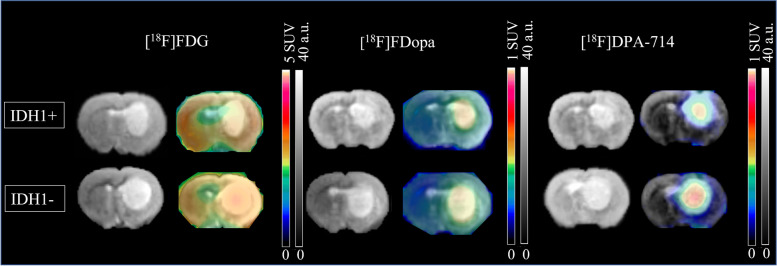
In vitro, IDH1+ cells (i.e. cells expressing the IDH1 mutation) showed lower levels of [F]DPA-714 uptake compared to IDH1- cells (p < 0.01). These results were confirmed in vivo with lower [F]DPA-714 uptake in IDH+ tumors (3.90 versus 5.52 for TBR, p = 0.03). Different values of [F]DPA-714 and [F] FDopa RRT (respectively 11.07 versus 22.33 and 2.69 versus - 1.81 for IDH+ and IDH- tumors, p < 0.02) were also observed between the two types of tumors. RRT [F]DPA-714 provided the best diagnostic performance to discriminate between the two cell lines (AUC of 100%, p < 0.01). Immuno-histological analyses revealed lower expression of Iba-1 and TSPO antibodies in IDH1+ tumors.
[18F]DPA-714 and [18F] FDopa both correlate with the presence of the IDH1 mutation in HGG. These radiotracers are therefore good candidates for translational studies investigating their clinical applications in patients.
本转化研究通过多示踪剂正电子发射断层扫描(PET)成像,来无创检测 IDH1 突变,该突变是神经胶质瘤的一个阳性预后因素。
利用 CRISP/Cas9 方法构建具有或不具有 IDH1 突变的 U87 人高级别神经胶质瘤(HGG)同基因细胞系,并对其进行立体定向移植到大鼠脑内,进行体外、体内和离体检测。使用针对糖酵解代谢([F]FDG)、氨基酸代谢([F]FDopa)和炎症([F]DPA-714)的放射性示踪剂,连续进行 3-4 天的 PET 成像。体外放射性示踪剂摄取用每百万细胞的百分比表示。对于体内检查的每种放射性示踪剂,静态分析包括最大和平均肿瘤与背景比(TBR 和 TBR)和代谢肿瘤体积(MTV)。动态分析包括从参考 Logan 模型提取的分布容积比(DVR)和相对滞留时间(RRT)。离体分析包括免疫分析。
体外,IDH1+细胞(即表达 IDH1 突变的细胞)与 IDH1-细胞相比,[F]DPA-714 摄取水平较低(p<0.01)。这些结果在体内得到了证实,IDH+肿瘤的[F]DPA-714 摄取水平较低(3.90 对 5.52 为 TBR,p=0.03)。两种类型肿瘤之间还观察到 [F]DPA-714 和 [F]FDopa RRT 值不同(分别为 IDH+和 IDH-肿瘤 11.07 对 22.33 和 2.69 对-1.81,p<0.02)。RRT [F]DPA-714 提供了区分两种细胞系的最佳诊断性能(AUC 为 100%,p<0.01)。免疫组织化学分析显示 IDH1+肿瘤中 Iba-1 和 TSPO 抗体的表达较低。
[18F]DPA-714 和 [18F]FDopa 均与 HGG 中 IDH1 突变的存在相关。因此,这些示踪剂是研究其在患者中临床应用的转化研究的良好候选物。