Department of Radiology, Union Hospital, Tongji Medical College, Huazhong University of Science and Technology, Wuhan, Hubei, China.
Hubei Province Key Laboratory of Molecular Imaging, Wuhan, 430022, China.
J Cardiovasc Magn Reson. 2022 Mar 21;24(1):19. doi: 10.1186/s12968-022-00849-1.
Both stenosis rate and intraplaque hemorrhage (IPH) are important predictors of stroke risk. Simultaneous non-contrast angiography and intraplaque hemorrhage (SNAP) cardiovascular magnetic resonance (CMR) imaging can detect both stenosis rate and IPH. We aimed to evaluate consistency between SNAP and digital subtraction angiography (DSA) to assess symptomatic patients with stroke and explore the performance of SNAP to identify IPH and the clinical factors associated with IPH.
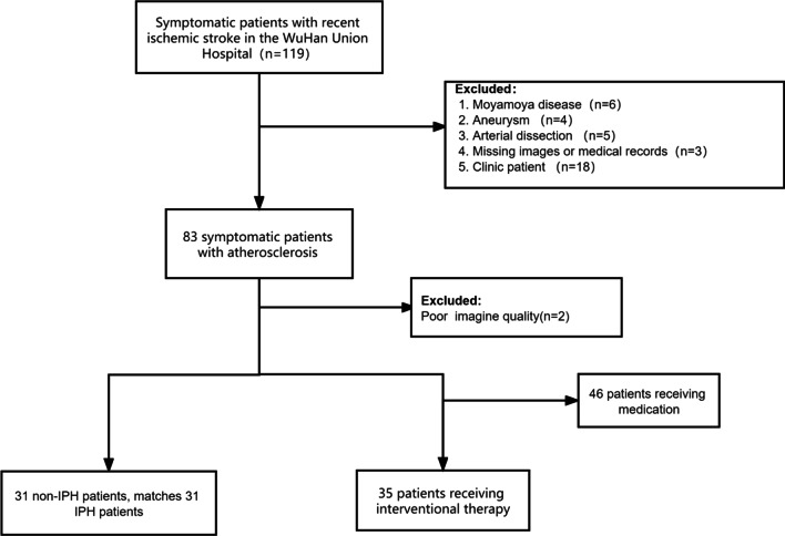
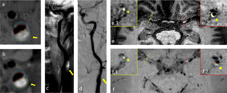
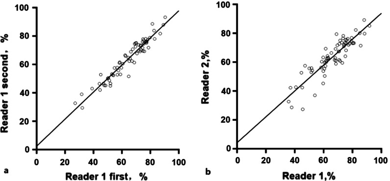
Eighty-one symptomatic patients with stroke, admitted to Wuhan Union Hospital who underwent CMR high-resolution vessel wall imaging (HR-VWI) and SNAP, were retrospectively identified. For patients who received interventional therapy, the imaging functions of SNAP and HR-VWI were compared with DSA. The diameters of the intracranial and carotid vessels were measured, and stenotic vessels were identified. The consistency of SNAP and HR-VWI in identifying IPH was also examined, and the correlations between IPH and clinical factors were analyzed.
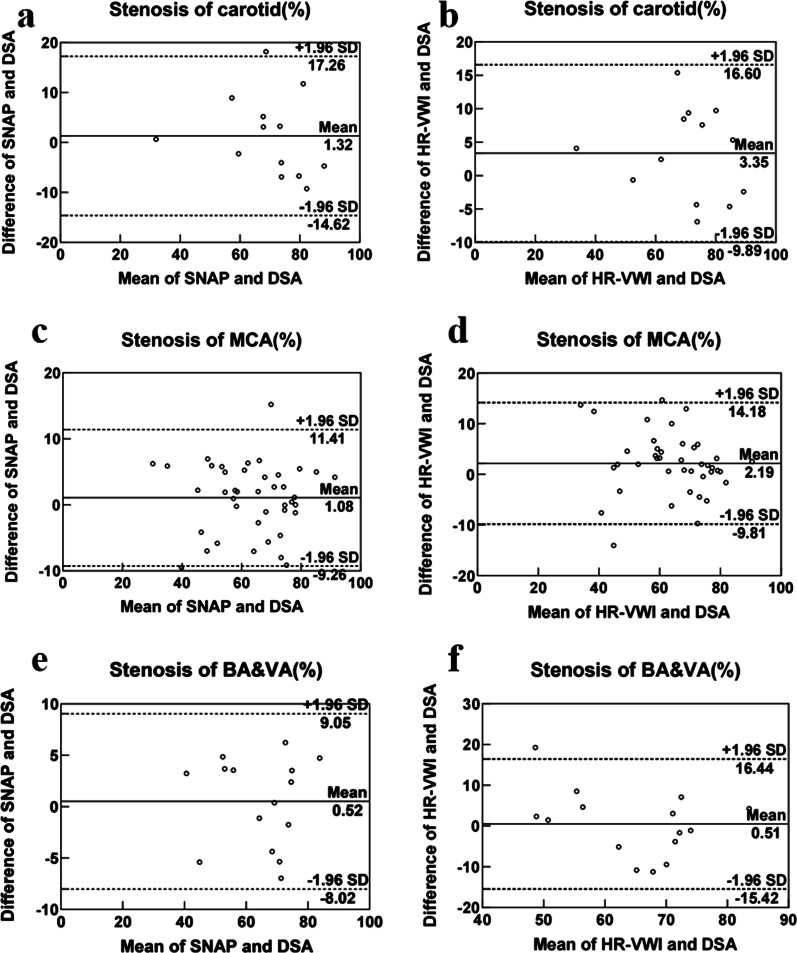
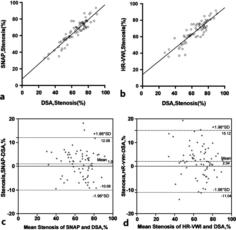
SNAP was more consistent with DSA than HR-VWI in measuring vascular stenosis (intraclass correlation coefficient [ICC] = 0.917, ICC = 0.878). Regarding the diameter measurements of each intracranial and carotid vessel segment, SNAP was superior or similar to HR-VWI, and both were consistent with DSA in the measurement of major intracranial vascular segments. HR-VWI and SNAP exhibited acceptable agreement in identifying IPH (Kappa = 0.839, 95% confidence interval [CI]: 0.704-0.974). Patients who underwent interventional therapy had a higher plaque burden (P < 0.001). Patients with IPH had lower levels of high-density lipoprotein cholesterol (HDL) (P = 0.038) and higher levels of blood glucose (P = 0.007) and cystatin C (P = 0.040).
CMR SNAP is consistent with DSA in measuring vessel diameters and identifying atherosclerosis stenosis in each intracranial and carotid vessel segment. SNAP is also a potential alternative to HR-VWI in identifying stenosis and IPH.
狭窄率和斑块内出血(IPH)都是中风风险的重要预测指标。同时进行非对比血管造影和斑块内出血(SNAP)心血管磁共振(CMR)成像可以同时检测狭窄率和 IPH。我们旨在评估 SNAP 与数字减影血管造影(DSA)之间的一致性,以评估有中风症状的患者,并探讨 SNAP 识别 IPH 的性能以及与 IPH 相关的临床因素。
回顾性纳入 81 例因中风入住武汉协和医院并接受 CMR 高分辨率血管壁成像(HR-VWI)和 SNAP 的症状性中风患者。对于接受介入治疗的患者,比较了 SNAP 和 HR-VWI 的成像功能。测量颅内和颈动脉血管的直径,并识别狭窄血管。还检查了 SNAP 和 HR-VWI 识别 IPH 的一致性,并分析了 IPH 与临床因素的相关性。
SNAP 在测量血管狭窄方面比 HR-VWI 更与 DSA 一致(组内相关系数 [ICC] = 0.917,ICC = 0.878)。对于每个颅内和颈动脉血管节段的直径测量,SNAP 优于或类似于 HR-VWI,并且在主要颅内血管节段的测量方面与 DSA 一致。HR-VWI 和 SNAP 在识别 IPH 方面具有可接受的一致性(Kappa = 0.839,95%置信区间 [CI]:0.704-0.974)。接受介入治疗的患者斑块负荷更高(P < 0.001)。IPH 患者的高密度脂蛋白胆固醇(HDL)水平较低(P = 0.038),血糖(P = 0.007)和胱抑素 C(P = 0.040)水平较高。
CMR SNAP 在测量血管直径和识别每个颅内和颈动脉血管节段的动脉粥样硬化狭窄方面与 DSA 一致。SNAP 也是识别狭窄和 IPH 的 HR-VWI 的潜在替代方法。