Department of Radiology, Tongji Hospital, Huazhong University of Science and Technology, Wuhan, China.
Department of Radiology, University of Washington, Seattle, Washington, USA.
J Magn Reson Imaging. 2017 Oct;46(4):1045-1052. doi: 10.1002/jmri.25653. Epub 2017 Feb 6.
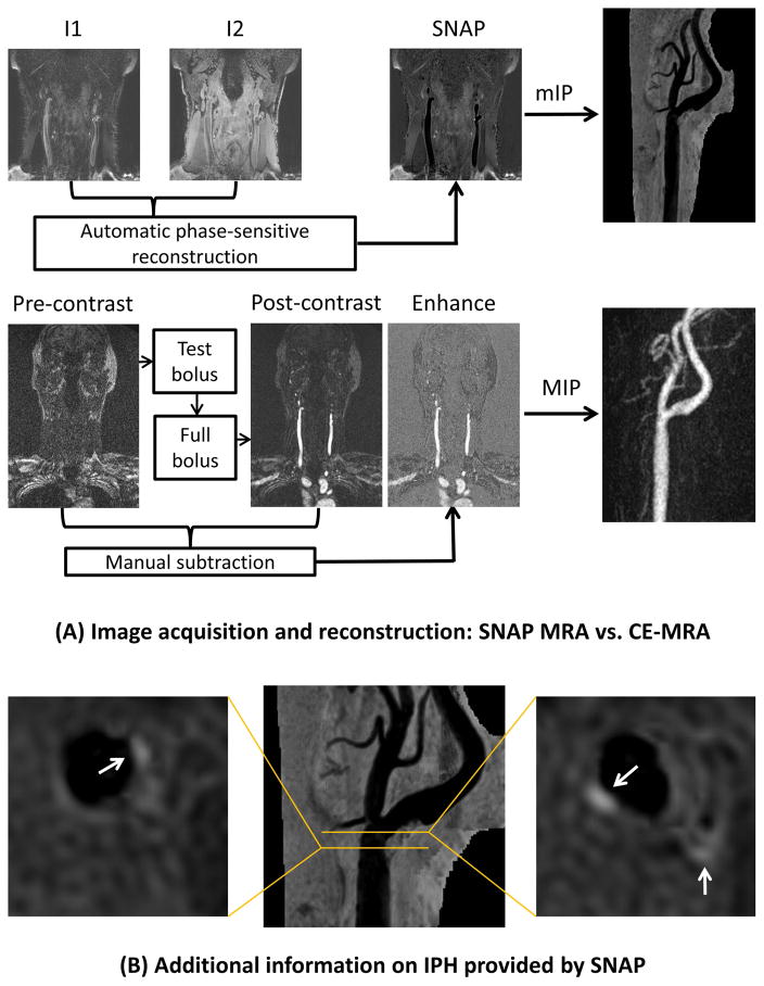
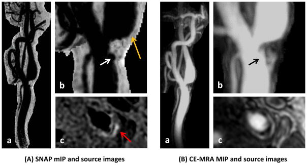
To evaluate in a proof-of-concept study the feasibility of Simultaneous Noncontrast Angiography and intraPlaque hemorrhage (SNAP) imaging as a clinical magnetic resonance angiography (MRA) technique for measuring carotid stenosis. There is a growing interest in detecting intraplaque hemorrhage (IPH) during the clinical management of carotid disease, yet luminal stenosis has remained indispensable during clinical decision-making. SNAP imaging has been proposed as a novel IPH imaging technique that provides carotid MRA with no added scan time. Flowing blood shows negative signal on SNAP because of phase-sensitive inversion recovery.
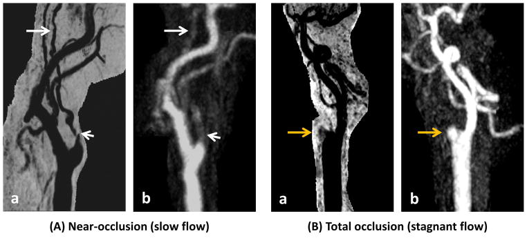
In all, 58 asymptomatic subjects with 16-79% stenosis on ultrasound were scanned at 3T by SNAP with 0.8 mm isotropic resolution and 16 cm longitudinal coverage. Two readers measured luminal stenosis of bilateral carotid arteries (n = 116) on minimum intensity projections of SNAP using the NASCET criteria. In the subset (48 arteries) with contrast-enhanced (CE) MRA available for comparison, luminal stenosis was also measured on maximum intensity projections of CE-MRA.
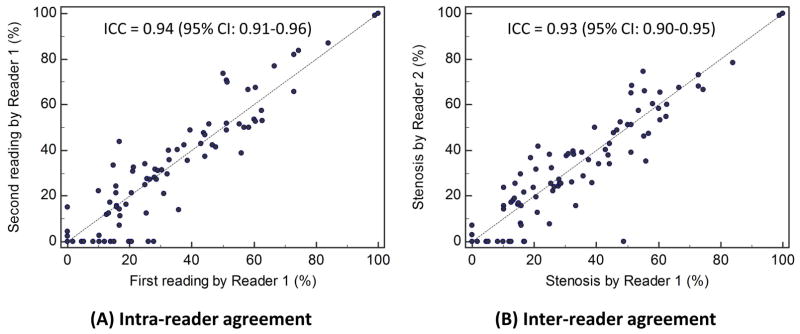
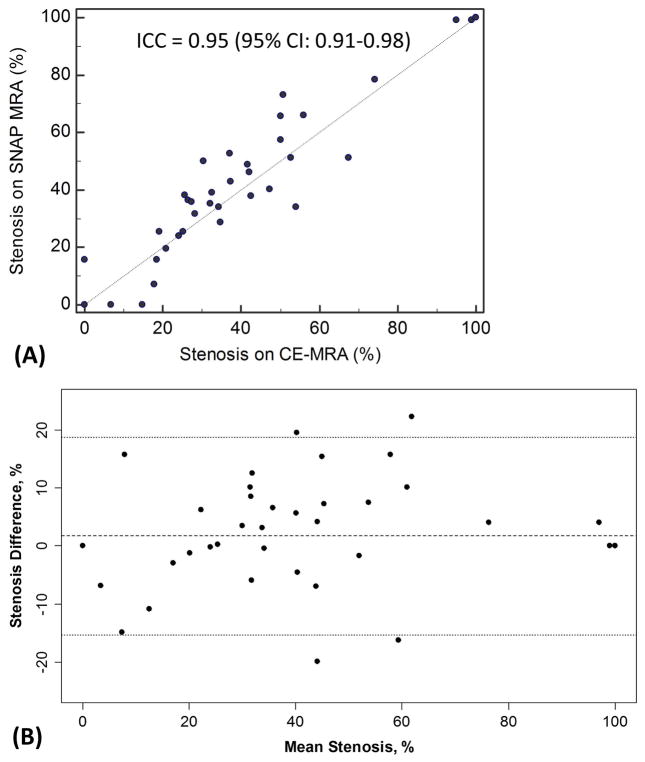
Intraclass correlation coefficients (ICCs) with 95% confidence intervals were 0.94 (0.90-0.96) and 0.93 (0.88-0.96) for intra- and interreader agreement on stenosis measurements, respectively. Corresponding kappas for grading stenosis (0-29%, 30-69%, 70-99%, and 100%) were 0.79 (0.67-0.89) and 0.80 (0.68-0.90). Agreement between SNAP and CE-MRA was high (ICC: 0.95 [0.90-0.98]; kappa: 0.82 [0.71-0.93]).
As a dedicated IPH-imaging sequence, SNAP also provided carotid stenosis measurement that showed high intra- and interreader consistency and excellent agreement with CE-MRA. Further comparisons with digital subtraction angiography and other noninvasive techniques are warranted.
2 Technical Efficacy: Stage 2 J. Magn. Reson. Imaging 2017;46:1045-1052.
在一项概念验证研究中评估 Simultaneous Noncontrast Angiography 和 intraPlaque hemorrhage(SNAP)成像作为一种临床磁共振血管造影(MRA)技术用于测量颈动脉狭窄的可行性。在颈动脉疾病的临床管理中,检测斑块内出血(IPH)的兴趣日益浓厚,但在临床决策中仍然需要管腔狭窄。SNAP 成像已被提议作为一种新的 IPH 成像技术,可为颈动脉 MRA 提供无额外扫描时间。由于相位敏感反转恢复,流动血液在 SNAP 上显示负信号。
共有 58 名无症状受试者,超声显示狭窄程度为 16%-79%,在 3T 上进行 SNAP 扫描,具有 0.8mm 各向同性分辨率和 16cm 纵向覆盖范围。两位读者使用 NASCET 标准在 SNAP 的最小强度投影上测量双侧颈动脉的管腔狭窄(n=116)。在具有对比增强(CE)MRA 可供比较的亚组(48 个动脉)中,也在 CE-MRA 的最大强度投影上测量管腔狭窄。
狭窄程度测量的组内和组间一致性的 intraclass 相关系数(ICC)分别为 0.94(0.90-0.96)和 0.93(0.88-0.96)。分级狭窄(0-29%、30-69%、70-99%和 100%)的相应kappa 值为 0.79(0.67-0.89)和 0.80(0.68-0.90)。SNAP 与 CE-MRA 之间的一致性很高(ICC:0.95[0.90-0.98];kappa:0.82[0.71-0.93])。
作为一种专用的 IPH 成像序列,SNAP 还提供了颈动脉狭窄测量,具有高的组内和组间一致性,并与 CE-MRA 具有极好的一致性。需要与数字减影血管造影和其他非侵入性技术进行进一步比较。
2 技术功效:第 2 阶段 J. Magn. Reson. Imaging 2017;46:1045-1052.