• 文献检索
  • 文档翻译
  • 深度研究
  • 学术资讯
  • Suppr Zotero 插件Zotero 插件
  • 邀请有礼
  • 套餐&价格
  • 历史记录
应用&插件
Suppr Zotero 插件Zotero 插件浏览器插件Mac 客户端Windows 客户端微信小程序
定价
高级版会员购买积分包购买API积分包
服务
文献检索文档翻译深度研究API 文档MCP 服务
关于我们
关于 Suppr公司介绍联系我们用户协议隐私条款
关注我们

Suppr 超能文献

核心技术专利:CN118964589B侵权必究
粤ICP备2023148730 号-1Suppr @ 2026

文献检索

告别复杂PubMed语法,用中文像聊天一样搜索,搜遍4000万医学文献。AI智能推荐,让科研检索更轻松。

立即免费搜索

文件翻译

保留排版,准确专业,支持PDF/Word/PPT等文件格式,支持 12+语言互译。

免费翻译文档

深度研究

AI帮你快速写综述,25分钟生成高质量综述,智能提取关键信息,辅助科研写作。

立即免费体验

1.5T 和 3T MRI 中人工耳蜗引起的伪影的尺寸和形态。

Dimensions and forms of artefacts in 1.5 T and 3 T MRI caused by cochlear implants.

机构信息

Department of Otorhinolaryngology, Head and Neck Surgery, Medical University of Innsbruck, Anichstr. 35, 6020, Innsbruck, Tyrol, Austria.

Department of Radiology, Medical University of Innsbruck, Anichstr. 35, 6020, Innsbruck, Tyrol, Austria.

出版信息

Sci Rep. 2022 Mar 22;12(1):4884. doi: 10.1038/s41598-022-08988-2.

DOI:10.1038/s41598-022-08988-2
PMID:35318407
原文链接:https://pmc.ncbi.nlm.nih.gov/articles/PMC8940987/
Abstract

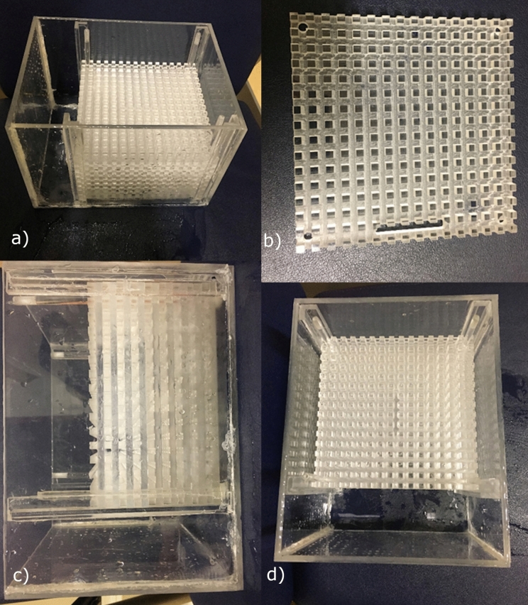
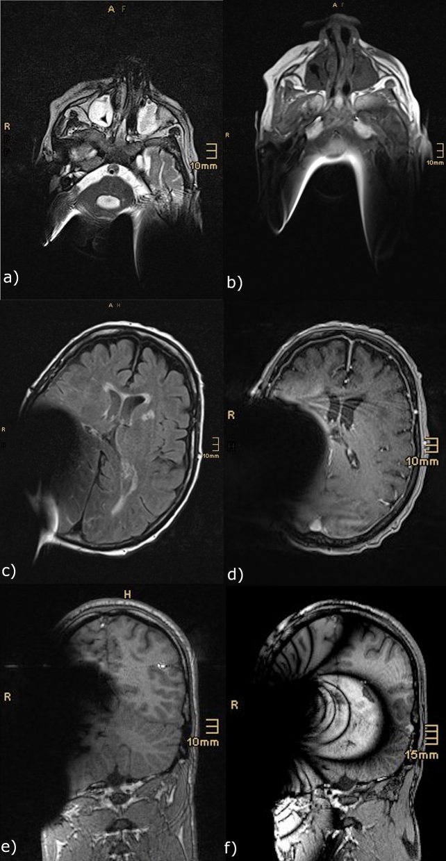
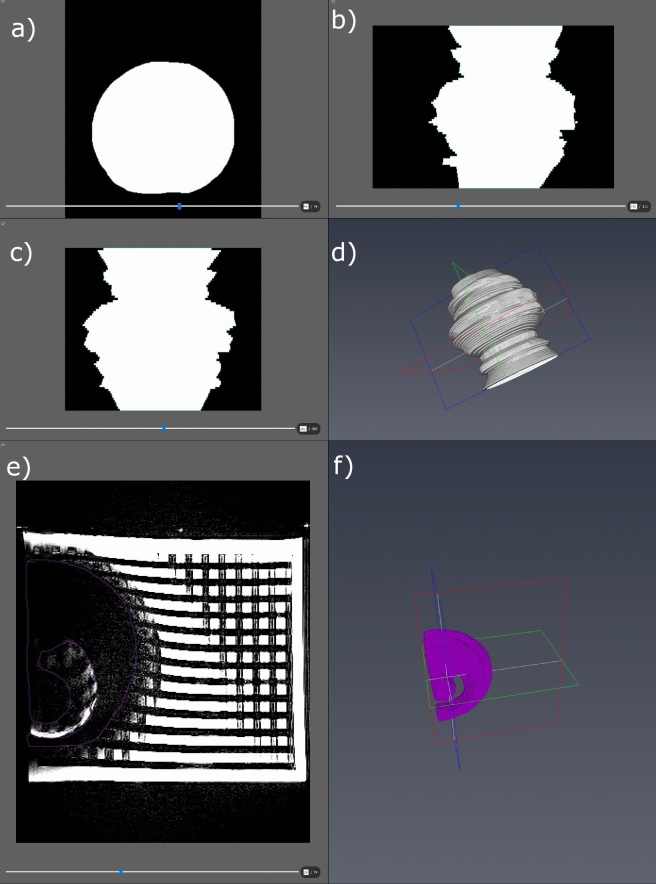
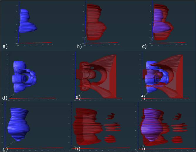
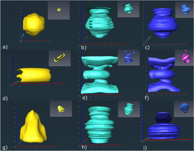
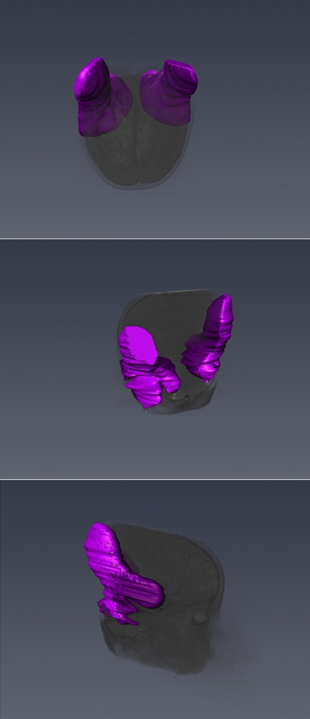
Cochlear implantation is a standard treatment option due to expanding indications. Cranial magnetic resonance imaging (cMRI) has become a widespread diagnostic tool. Therefore, an increased number of cochlear implant (CI) users are undergoing cMRI scans. This study aimed to investigate the issue of the CI magnet impacting MRI quality and artifacts. 1.5 T and 3 T MRI scans with 4 defined sequences (T2-TSE, T2-TIRM, T1-3D-MPRAGE, and TDI) were performed on a phantom with a CI (SYNCHRONY System by MED-EL Austria) in place. The resulting MRI artifacts were retrospectively compared to MRI artifacts observed in patients with a CI. All images were transferred to AMIRA and visualized by manual segmentation. Usable image quality was achieved in three sequences (T2-TSE, T2-TIRM and T1-mprage). Observed artifacts differed in shape and size depending on the sequence. Maximum diameters of signal void areas ranged from 58 × 108 × 98 mm to 127 × 123 × 153 mm. Image distortions were larger. MRI artifacts caused by the SYNCHRONY system are asymmetric with varying shape, depending on the sequence. The phantom artefacts are similar to those in CI users. Considering the observed asymmetry, the hypothesis of varying implantation locations resulting in varying positions of the signal void area needs to be further investigated.

摘要

人工耳蜗植入术是一种标准的治疗选择,因为适应证在不断扩大。颅磁共振成像(cMRI)已成为一种广泛使用的诊断工具。因此,越来越多的人工耳蜗植入(CI)使用者接受 cMRI 扫描。本研究旨在探讨 CI 磁铁对 MRI 质量和伪影的影响问题。在一个带有 CI(由 MED-EL 奥地利公司生产的 SYNCHRONY 系统)的体模上进行了 1.5T 和 3T 的 MRI 扫描,共 4 个定义序列(T2-TSE、T2-TIRM、T1-3D-MPRAGE 和 TDI)。将得到的 MRI 伪影与 CI 患者的 MRI 伪影进行回顾性比较。所有图像都被转移到 AMIRA 并通过手动分割进行可视化。在三个序列(T2-TSE、T2-TIRM 和 T1-MPRAGE)中获得了可用的图像质量。观察到的伪影的形状和大小因序列而异。信号缺失区域的最大直径范围从 58×108×98mm 到 127×123×153mm。图像失真更大。SYNCHRONY 系统引起的 MRI 伪影不对称,形状各异,取决于序列。体模伪影与 CI 用户的相似。考虑到观察到的不对称性,需要进一步研究不同植入位置导致信号缺失区域位置不同的假设。

https://cdn.ncbi.nlm.nih.gov/pmc/blobs/eaab/8940987/377dae808e6f/41598_2022_8988_Fig6_HTML.jpg
https://cdn.ncbi.nlm.nih.gov/pmc/blobs/eaab/8940987/d71f5752e3ee/41598_2022_8988_Fig1_HTML.jpg
https://cdn.ncbi.nlm.nih.gov/pmc/blobs/eaab/8940987/f308e9c97237/41598_2022_8988_Fig2_HTML.jpg
https://cdn.ncbi.nlm.nih.gov/pmc/blobs/eaab/8940987/562ac78c0999/41598_2022_8988_Fig3_HTML.jpg
https://cdn.ncbi.nlm.nih.gov/pmc/blobs/eaab/8940987/b777c88c0c1e/41598_2022_8988_Fig4_HTML.jpg
https://cdn.ncbi.nlm.nih.gov/pmc/blobs/eaab/8940987/d411712f74be/41598_2022_8988_Fig5_HTML.jpg
https://cdn.ncbi.nlm.nih.gov/pmc/blobs/eaab/8940987/377dae808e6f/41598_2022_8988_Fig6_HTML.jpg

相似文献

1
Dimensions and forms of artefacts in 1.5 T and 3 T MRI caused by cochlear implants.1.5T 和 3T MRI 中人工耳蜗引起的伪影的尺寸和形态。
Sci Rep. 2022 Mar 22;12(1):4884. doi: 10.1038/s41598-022-08988-2.
2
Dimensions of artefacts caused by cochlear and auditory brainstem implants in magnetic resonance imaging.人工耳蜗和听脑干植入物在磁共振成像中的伪影维度。
Cochlear Implants Int. 2020 Mar;21(2):67-74. doi: 10.1080/14670100.2019.1668617. Epub 2019 Sep 25.
3
MRI-induced artifact by a cochlear implant with a novel magnet system: an experimental cadaver study.新型磁体系统的人工耳蜗引起的 MRI 伪影:一项实验性尸体研究。
Eur Arch Otorhinolaryngol. 2021 Oct;278(10):3753-3762. doi: 10.1007/s00405-020-06464-z. Epub 2020 Nov 18.
4
Intraindividual Comparison of Image Artifacts of Two Generations of Rotatable Cochlear Implant Magnets.两代可旋转人工耳蜗磁铁的个体内图像伪影比较。
J Int Adv Otol. 2024 Jul 29;20(4):306-311. doi: 10.5152/iao.2024.231466.
5
MRI Artifacts and Cochlear Implant Positioning at 3 T In Vivo.3T 磁共振成像伪影与体内人工耳蜗植入位置
Otol Neurotol. 2015 Jul;36(6):972-6. doi: 10.1097/MAO.0000000000000720.
6
Feasibility and limitations of head MRI in patients with cochlear implants.人工耳蜗植入患者行头部 MRI 的可行性和局限性。
Auris Nasus Larynx. 2023 Dec;50(6):874-879. doi: 10.1016/j.anl.2023.04.001. Epub 2023 Apr 17.
7
Cranial MRI in a young child with cochlear implants after bilateral magnet removal.双侧磁铁取出后植入人工耳蜗的幼儿的头颅磁共振成像
Int J Pediatr Otorhinolaryngol. 2017 Dec;103:1-4. doi: 10.1016/j.ijporl.2017.09.028. Epub 2017 Sep 28.
8
Parametric Assessment of the Effect of Cochlear Implant Positioning on Brain MRI Artefacts at 3 T.人工耳蜗植入位置对 3T 脑 MRI 伪影影响的参数评估。
Otol Neurotol. 2021 Dec 1;42(10):e1449-e1456. doi: 10.1097/MAO.0000000000003281.
9
Characterizing Cochlear Implant Magnet-Related MRI Artifact.描述人工耳蜗磁铁相关的 MRI 伪影。
Ann Otol Rhinol Laryngol. 2023 Mar;132(3):250-258. doi: 10.1177/00034894221089143. Epub 2022 Apr 5.
10
Effect of Head Position and Magnetic Resonance Sequence on Cochlear Implant-Related Artifact Size and Internal Auditory Canal Visibility.头位和磁共振序列对人工耳蜗植入相关伪影大小及内听道显示的影响。
Otol Neurotol. 2023 Feb 1;44(2):e73-e80. doi: 10.1097/MAO.0000000000003738.

本文引用的文献

1
Parametric Assessment of the Effect of Cochlear Implant Positioning on Brain MRI Artefacts at 3 T.人工耳蜗植入位置对 3T 脑 MRI 伪影影响的参数评估。
Otol Neurotol. 2021 Dec 1;42(10):e1449-e1456. doi: 10.1097/MAO.0000000000003281.
2
MRI-induced artifact by a cochlear implant with a novel magnet system: an experimental cadaver study.新型磁体系统的人工耳蜗引起的 MRI 伪影:一项实验性尸体研究。
Eur Arch Otorhinolaryngol. 2021 Oct;278(10):3753-3762. doi: 10.1007/s00405-020-06464-z. Epub 2020 Nov 18.
3
Effect of head position on cochlear implant MRI artifact.
头位对人工耳蜗 MRI 伪影的影响。
Eur Arch Otorhinolaryngol. 2021 Aug;278(8):2763-2767. doi: 10.1007/s00405-020-06349-1. Epub 2020 Sep 9.
4
Comparison of Cochlear Implant Magnets and Their MRI Artifact Size.人工耳蜗磁铁的比较及其磁共振成像伪影大小。
Biomed Res Int. 2020 Jan 10;2020:5086291. doi: 10.1155/2020/5086291. eCollection 2020.
5
Magnetic Resonance Imaging Artifacts and Cochlear Implant Positioning at 1.5 T In Vivo.1.5T 体内磁共振成像伪影与人工耳蜗植入位置
Biomed Res Int. 2018 Nov 8;2018:9163285. doi: 10.1155/2018/9163285. eCollection 2018.
6
[MRI in patients with auditory implants equipped with implanted magnets-an update : Overview and procedural management].[配备植入式磁体的听觉植入患者的MRI——最新进展:概述与程序管理]
Radiologe. 2019 Jan;59(1):48-56. doi: 10.1007/s00117-018-0462-9.
7
Non-contrast magnetic resonance imaging for monitoring patients with acoustic neuroma.用于监测听神经瘤患者的非增强磁共振成像
J Laryngol Otol. 2018 Sep;132(9):780-785. doi: 10.1017/S0022215118001342. Epub 2018 Aug 17.
8
Visualization of the Membranous Labyrinth and Nerve Fiber Pathways in Human and Animal Inner Ears Using MicroCT Imaging.使用显微计算机断层扫描成像技术可视化人类和动物内耳中的膜迷路和神经纤维通路。
Front Neurosci. 2018 Jul 31;12:501. doi: 10.3389/fnins.2018.00501. eCollection 2018.
9
Time window and "tissue window": two approaches to assist decision-making in strokes.时间窗和“组织窗”:两种辅助脑卒中决策的方法。
J Neurol. 2019 Feb;266(2):283-288. doi: 10.1007/s00415-018-8933-5. Epub 2018 Jun 19.
10
Meta-analysis of computed tomography angiography versus magnetic resonance angiography for intracranial aneurysm.计算机断层扫描血管造影术与磁共振血管造影术用于颅内动脉瘤的荟萃分析
Medicine (Baltimore). 2018 May;97(20):e10771. doi: 10.1097/MD.0000000000010771.