• 文献检索
  • 文档翻译
  • 深度研究
  • 学术资讯
  • Suppr Zotero 插件Zotero 插件
  • 邀请有礼
  • 套餐&价格
  • 历史记录
应用&插件
Suppr Zotero 插件Zotero 插件浏览器插件Mac 客户端Windows 客户端微信小程序
定价
高级版会员购买积分包购买API积分包
服务
文献检索文档翻译深度研究API 文档MCP 服务
关于我们
关于 Suppr公司介绍联系我们用户协议隐私条款
关注我们

Suppr 超能文献

核心技术专利:CN118964589B侵权必究
粤ICP备2023148730 号-1Suppr @ 2026

文献检索

告别复杂PubMed语法,用中文像聊天一样搜索,搜遍4000万医学文献。AI智能推荐,让科研检索更轻松。

立即免费搜索

文件翻译

保留排版,准确专业,支持PDF/Word/PPT等文件格式,支持 12+语言互译。

免费翻译文档

深度研究

AI帮你快速写综述,25分钟生成高质量综述,智能提取关键信息,辅助科研写作。

立即免费体验

使用显微计算机断层扫描成像技术可视化人类和动物内耳中的膜迷路和神经纤维通路。

Visualization of the Membranous Labyrinth and Nerve Fiber Pathways in Human and Animal Inner Ears Using MicroCT Imaging.

作者信息

Glueckert Rudolf, Johnson Chacko Lejo, Schmidbauer Dominik, Potrusil Thomas, Pechriggl Elisabeth J, Hoermann Romed, Brenner Erich, Reka Alen, Schrott-Fischer Anneliese, Handschuh Stephan

机构信息

Department of Otolaryngology, Medical University of Innsbruck, Innsbruck, Austria.

University Clinics Innsbruck, Tirol Kliniken, University Clinic for Ear, Nose and Throat Medicine Innsbruck, Innsbruck, Austria.

出版信息

Front Neurosci. 2018 Jul 31;12:501. doi: 10.3389/fnins.2018.00501. eCollection 2018.

DOI:10.3389/fnins.2018.00501
PMID:30108474
原文链接:https://pmc.ncbi.nlm.nih.gov/articles/PMC6079228/
Abstract

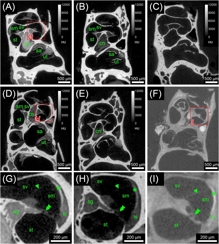
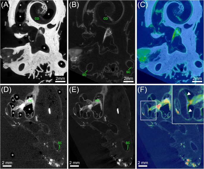
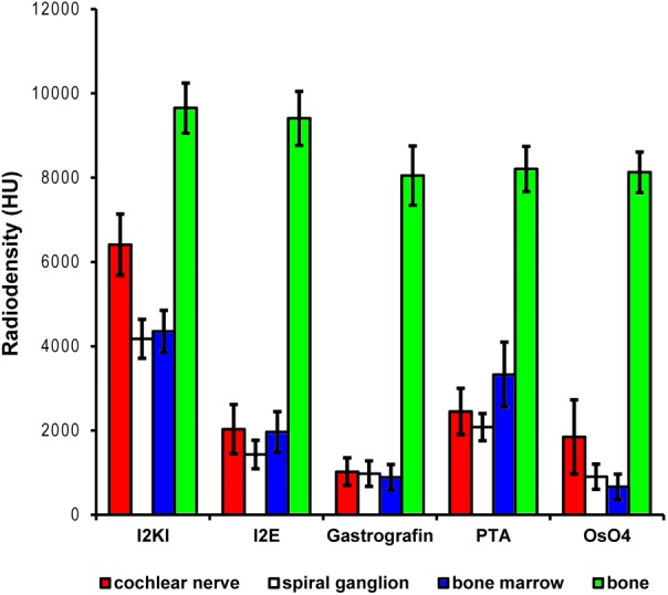
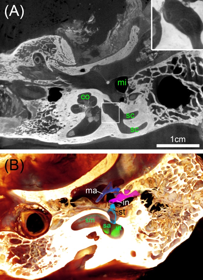
Design and implantation of bionic implants for restoring impaired hair cell function relies on accurate knowledge about the microanatomy and nerve fiber pathways of the human inner ear and its variation. Non-destructive isotropic imaging of soft tissues of the inner ear with lab-based microscopic X-ray computed tomography (microCT) offers high resolution but requires contrast enhancement using compounds with high X-ray attenuation. We evaluated different contrast enhancement techniques in mice, cat, and human temporal bones to differentially visualize the membranous labyrinth, sensory epithelia, and their innervating nerves together with the facial nerve and middle ear. Lugol's iodine potassium iodine (IKI) gave high soft tissue contrast in ossified specimens but failed to provide unambiguous identification of smaller nerve fiber bundles inside small bony canals. Fixation or post-fixation with osmium tetroxide followed by decalcification in EDTA provided superior contrast for nerve fibers and membranous structures. We processed 50 human temporal bones and acquired microCT scans with 15 μm voxel size. Subsequently we segmented sensorineural structures and the endolymphatic compartment for 3D representations to serve for morphometric variation analysis. We tested higher resolution image acquisition down to 3.0 μm voxel size in human and 0.5 μm in mice, which provided a unique level of detail and enabled us to visualize single neurons and hair cells in the mouse inner ear, which could offer an alternative quantitative analysis of cell numbers in smaller animals. Bigger ossified human temporal bones comprising the middle ear and mastoid bone can be contrasted with IKI and imaged in toto at 25 μm voxel size. These data are suitable for surgical planning for electrode prototype placements. A preliminary assessment of geometric changes through tissue processing resulted in 1.6% volume increase caused during decalcification by EDTA and 0.5% volume increase caused by partial dehydration to 70% ethanol, which proved to be the best mounting medium for microCT image acquisition.

摘要

用于恢复受损毛细胞功能的仿生植入物的设计与植入依赖于对人类内耳及其变异的微观解剖结构和神经纤维通路的准确了解。基于实验室的微观X射线计算机断层扫描(microCT)对内耳软组织进行无损各向同性成像可提供高分辨率,但需要使用具有高X射线衰减的化合物进行对比增强。我们在小鼠、猫和人类颞骨中评估了不同的对比增强技术,以分别可视化膜迷路、感觉上皮及其支配神经以及面神经和中耳。卢戈氏碘钾碘(IKI)在骨化标本中提供了高软组织对比度,但未能明确识别小骨管内较小的神经纤维束。用四氧化锇固定或后固定,然后在乙二胺四乙酸(EDTA)中脱钙,可为神经纤维和膜结构提供更好的对比度。我们处理了50块人类颞骨,并以15μm的体素大小获取了microCT扫描图像。随后,我们对感觉神经结构和内淋巴腔进行分割以进行三维重建,用于形态计量变异分析。我们测试了更高分辨率的图像采集,人类的体素大小低至3.0μm,小鼠的低至0.5μm,这提供了独特的细节水平,使我们能够在小鼠内耳中可视化单个神经元和毛细胞,这可为较小动物中的细胞数量提供另一种定量分析方法。包含中耳和乳突骨的较大骨化人类颞骨可用IKI进行对比,并以25μm的体素大小进行整体成像。这些数据适用于电极原型放置的手术规划。通过组织处理对几何变化的初步评估表明,EDTA脱钙过程中导致体积增加了1.6%,部分脱水至70%乙醇导致体积增加了0.5%,70%乙醇被证明是microCT图像采集的最佳固定介质。

https://cdn.ncbi.nlm.nih.gov/pmc/blobs/c36a/6079228/1acb9daf2afa/fnins-12-00501-g009.jpg
https://cdn.ncbi.nlm.nih.gov/pmc/blobs/c36a/6079228/ee19c5efbb7e/fnins-12-00501-g001.jpg
https://cdn.ncbi.nlm.nih.gov/pmc/blobs/c36a/6079228/815becab9402/fnins-12-00501-g002.jpg
https://cdn.ncbi.nlm.nih.gov/pmc/blobs/c36a/6079228/abd6cdcbc04f/fnins-12-00501-g003.jpg
https://cdn.ncbi.nlm.nih.gov/pmc/blobs/c36a/6079228/ab1cd7dc589d/fnins-12-00501-g004.jpg
https://cdn.ncbi.nlm.nih.gov/pmc/blobs/c36a/6079228/57275402a14a/fnins-12-00501-g005.jpg
https://cdn.ncbi.nlm.nih.gov/pmc/blobs/c36a/6079228/4fe4b3bb2b7d/fnins-12-00501-g006.jpg
https://cdn.ncbi.nlm.nih.gov/pmc/blobs/c36a/6079228/aa98f1d8cbf4/fnins-12-00501-g007.jpg
https://cdn.ncbi.nlm.nih.gov/pmc/blobs/c36a/6079228/2868bad0dcc9/fnins-12-00501-g008.jpg
https://cdn.ncbi.nlm.nih.gov/pmc/blobs/c36a/6079228/1acb9daf2afa/fnins-12-00501-g009.jpg

相似文献

1
Visualization of the Membranous Labyrinth and Nerve Fiber Pathways in Human and Animal Inner Ears Using MicroCT Imaging.使用显微计算机断层扫描成像技术可视化人类和动物内耳中的膜迷路和神经纤维通路。
Front Neurosci. 2018 Jul 31;12:501. doi: 10.3389/fnins.2018.00501. eCollection 2018.
2
Analysis of Vestibular Labyrinthine Geometry and Variation in the Human Temporal Bone.人类颞骨前庭迷路的几何形状及变异分析
Front Neurosci. 2018 Feb 26;12:107. doi: 10.3389/fnins.2018.00107. eCollection 2018.
3
Volumetric atlas of the rat inner ear from microCT and iDISCO+ cleared temporal bones.基于微计算机断层扫描(microCT)和免疫组织化学成像系统(iDISCO+)清除颞骨构建的大鼠内耳体积图谱。
PeerJ. 2025 May 26;13:e19512. doi: 10.7717/peerj.19512. eCollection 2025.
4
Optimization of 3D-Visualization of Micro-Anatomical Structures of the Human Inner Ear in Osmium Tetroxide Contrast Enhanced Micro-CT Scans.锇酸增强显微CT扫描中人类内耳微观解剖结构的三维可视化优化
Front Neuroanat. 2018 May 22;12:41. doi: 10.3389/fnana.2018.00041. eCollection 2018.
5
Experimental Fusion of Contrast Enhanced High-Field Magnetic Resonance Imaging and High-Resolution Micro-Computed Tomography in Imaging the Mouse Inner Ear.对比增强高场磁共振成像与高分辨率微型计算机断层扫描在小鼠内耳成像中的实验融合
Open Neuroimag J. 2015 Jul 31;9:7-12. doi: 10.2174/1874440001509010007. eCollection 2015.
6
MicroCT for comparative morphology: simple staining methods allow high-contrast 3D imaging of diverse non-mineralized animal tissues.用于比较形态学的显微CT:简单染色方法可实现多种非矿化动物组织的高对比度三维成像。
BMC Physiol. 2009 Jun 22;9:11. doi: 10.1186/1472-6793-9-11.
7
Heavy metal staining, a comparative assessment of gadolinium chloride and osmium tetroxide for inner ear labyrinthine contrast enhancement using X-ray microtomography.重金属染色,使用X射线显微断层扫描对氯化钆和四氧化锇用于内耳迷路造影增强的比较评估。
Acta Otolaryngol. 2013 Jan;133(1):22-7. doi: 10.3109/00016489.2012.715751. Epub 2012 Sep 19.
8
Iodine potassium iodide improves the contrast-to-noise ratio of micro-computed tomography images of the human middle ear.碘化钾可提高人中耳微计算机断层扫描图像的对比度噪声比。
J Microsc. 2016 Dec;264(3):334-338. doi: 10.1111/jmi.12447. Epub 2016 Jul 21.
9
High-resolution T2-weighted MR imaging of the inner ear using a long echo-train-length 3D fast spin-echo sequence.使用长回波链长度三维快速自旋回波序列对内耳进行高分辨率T2加权磁共振成像。
Eur Radiol. 1996;6(3):369-74. doi: 10.1007/BF00180615.
10
High-resolution 7-Tesla magnetic resonance imaging and post-processing for 3-dimensional reconstruction of the membranous labyrinth in healthy adults.健康成年人膜迷路三维重建的高分辨率7特斯拉磁共振成像及后处理
Anat Rec (Hoboken). 2025 Mar 12. doi: 10.1002/ar.25647.

引用本文的文献

1
3D imaging of the human temporal bone by X-ray phase-contrast tomography.通过X射线相衬断层扫描对人类颞骨进行三维成像。
Npj Imaging. 2025 May 20;3(1):21. doi: 10.1038/s44303-025-00086-y.
2
The State of High-Resolution Imaging of the Human Inner Ear: A Look Into the Black Box.人类内耳的高分辨率成像现状:窥探黑匣子
Adv Sci (Weinh). 2025 Jul;12(28):e00556. doi: 10.1002/advs.202500556. Epub 2025 Jun 5.
3
Volume rendering technique and high-resolution microCT: 3D exploration of the cochlear anatomy.容积再现技术与高分辨率微型计算机断层扫描:耳蜗解剖结构的三维探索

本文引用的文献

1
Analysis of Vestibular Labyrinthine Geometry and Variation in the Human Temporal Bone.人类颞骨前庭迷路的几何形状及变异分析
Front Neurosci. 2018 Feb 26;12:107. doi: 10.3389/fnins.2018.00107. eCollection 2018.
2
Model-based Vestibular Afferent Stimulation: Modular Workflow for Analyzing Stimulation Scenarios in Patient Specific and Statistical Vestibular Anatomy.基于模型的前庭传入神经刺激:用于分析特定患者和统计性前庭解剖结构中刺激场景的模块化工作流程。
Front Neurosci. 2017 Dec 19;11:713. doi: 10.3389/fnins.2017.00713. eCollection 2017.
3
Find your way with X-Ray: Using microCT to correlate in vivo imaging with 3D electron microscopy.
Eur Arch Otorhinolaryngol. 2025 Apr 3. doi: 10.1007/s00405-025-09360-6.
4
3D virtual histology of rodent and primate cochleae with multi-scale phase-contrast X-ray tomography.利用多尺度相衬X射线断层扫描技术对啮齿动物和灵长类动物耳蜗进行3D虚拟组织学研究。
Sci Rep. 2025 Mar 7;15(1):7933. doi: 10.1038/s41598-025-89431-0.
5
Shared features in ear and kidney development - implications for oto-renal syndromes.耳肾发育的共同特征——对耳肾综合征的启示。
Dis Model Mech. 2024 Feb 1;17(2). doi: 10.1242/dmm.050447. Epub 2024 Feb 14.
6
Imaging of excised cochleae by micro-CT: staining, liquid embedding, and image modalities.切除的耳蜗的显微CT成像:染色、液体包埋及图像模式
J Med Imaging (Bellingham). 2023 Sep;10(5):053501. doi: 10.1117/1.JMI.10.5.053501. Epub 2023 Sep 25.
7
The Next Challenges of Vestibular Implantation in Humans.人类前庭植入的下一个挑战。
J Assoc Res Otolaryngol. 2023 Aug;24(4):401-412. doi: 10.1007/s10162-023-00906-1. Epub 2023 Jul 29.
8
Closing the Gap between the Auditory Nerve and Cochlear Implant Electrodes: Which Neurotrophin Cocktail Performs Best for Axonal Outgrowth and Is Electrical Stimulation Beneficial?弥合听神经与人工耳蜗电极之间的差距:哪种神经营养因子鸡尾酒在轴突生长方面表现最佳,电刺激是否有益?
Int J Mol Sci. 2023 Jan 19;24(3):2013. doi: 10.3390/ijms24032013.
9
Reversible contrast enhancement for visualization of human temporal bones using micro computed tomography.使用微型计算机断层扫描对人类颞骨进行可视化的可逆对比增强。
Front Surg. 2022 Oct 4;9:952348. doi: 10.3389/fsurg.2022.952348. eCollection 2022.
10
An optimized workflow for microCT imaging of formalin-fixed and paraffin-embedded (FFPE) early equine embryos.优化福尔马林固定石蜡包埋(FFPE)早期马胚胎 microCT 成像的工作流程。
Anat Histol Embryol. 2022 Sep;51(5):611-623. doi: 10.1111/ahe.12834. Epub 2022 Jul 18.
借助X射线找到方向:利用显微CT将体内成像与三维电子显微镜相关联。
Methods Cell Biol. 2017;140:277-301. doi: 10.1016/bs.mcb.2017.03.006. Epub 2017 Apr 21.
4
Automated analysis of human cochlea shape variability from segmented μCT images.基于分割 μCT 图像的人类耳蜗形态变异性的自动分析。
Comput Med Imaging Graph. 2017 Jul;59:1-12. doi: 10.1016/j.compmedimag.2017.04.002. Epub 2017 Apr 12.
5
Functional morphological adaptations of the bony labyrinth in marsupials (Mammalia, Theria).有袋类动物(哺乳纲,兽亚纲)骨迷路的功能形态学适应
J Morphol. 2017 Jun;278(6):742-749. doi: 10.1002/jmor.20669. Epub 2017 Mar 27.
6
Microscopic dual-energy CT (microDECT): a flexible tool for multichannel ex vivo 3D imaging of biological specimens.微观双能CT(microDECT):用于生物标本多通道离体三维成像的灵活工具。
J Microsc. 2017 Jul;267(1):3-26. doi: 10.1111/jmi.12543. Epub 2017 Mar 7.
7
Micro-CT versus synchrotron radiation phase contrast imaging of human cochlea.微计算机断层扫描与同步辐射相衬成像对人耳蜗的研究
J Microsc. 2017 Mar;265(3):349-357. doi: 10.1111/jmi.12507. Epub 2016 Dec 9.
8
Ultra-High-Field Magnetic Resonance Imaging of the Human Inner Ear at 11.7 Tesla.11.7特斯拉下人类内耳的超高场磁共振成像
Otol Neurotol. 2017 Jan;38(1):133-138. doi: 10.1097/MAO.0000000000001242.
9
Three-dimensional modeling of the cochlea by use of an arc fitting approach.使用圆弧拟合方法对耳蜗进行三维建模。
Comput Methods Biomech Biomed Engin. 2016 Dec;19(16):1785-1799. doi: 10.1080/10255842.2016.1188921. Epub 2016 Aug 9.
10
Cochlear implant-related three-dimensional characteristics determined by micro-computed tomography reconstruction.通过微计算机断层扫描重建确定的人工耳蜗相关三维特征
Clin Anat. 2017 Jan;30(1):39-43. doi: 10.1002/ca.22770. Epub 2016 Sep 13.