Discipline of Orthodontics, Faculty of Dentistry, the University of Hong Kong, Hong Kong SAR, China.
Department of Computer Science, Chu Hai College of Higher Education, Hong Kong SAR, China.
Clin Oral Investig. 2022 Jul;26(7):4947-4966. doi: 10.1007/s00784-022-04463-4. Epub 2022 Mar 23.
The present study aimed to determine the site and severity of maxillomandibular asymmetry before and after orthognathic surgery in asymmetric patients.
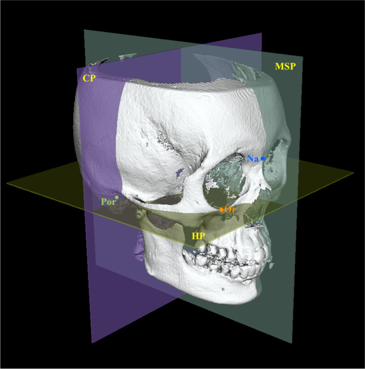
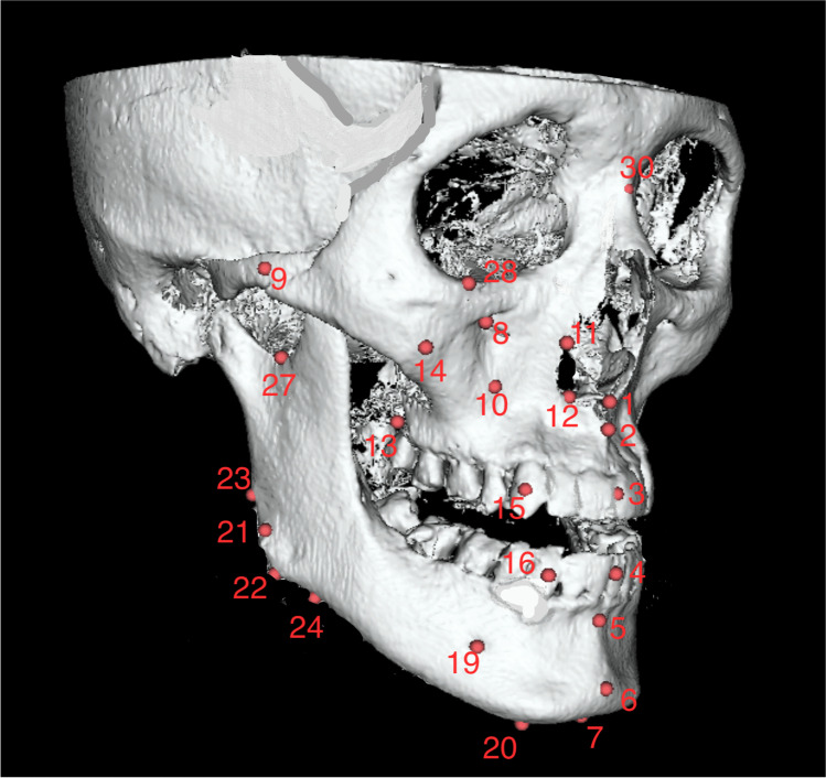
Presurgery and postsurgery cone beam computed tomography (CBCT) data of 21 facial asymmetry patients (7 males and 14 females, mean age: 23.0 ± 3.36 years) with soft tissue chin deviation ≥ 3 mm who had undergone bimaxillary surgery were evaluated. Seven midline and twenty bilateral hard tissue landmarks were identified for the evaluation of facial asymmetry and outcomes were assessed against age- and gender-matched control subjects.
In the asymmetry group, bilateral landmarks exhibited significant deviation in the mandible and midface regions. Before surgery, asymmetry was more severe at the mandibular midline and sites close to it, in the asymmetry group. Bimaxillary surgery proved to be highly effective, with a significant correction of the menton to a clinically normal value (2.90 mm, p < 0.001). After surgery, significant residual asymmetry was observed at the mental foramen (p = 0.001) in the R-L direction. Moreover, significant asymmetry persisted at the sigmoid notch (p = 0.001) in the S-I direction.
Mandibular midline landmarks and chin peripheral regions contribute significantly to overall facial asymmetry characteristics. Despite significant correction after bimaxillary surgery, asymmetry persisted at several sites, thereby requiring secondary correction. Comprehensive 3D presurgical planning is central for asymmetry correction in a single surgery.
The present study specifies the location of residual asymmetry sites and advocates the correction of those sites during initial surgery.
本研究旨在确定不对称患者正颌手术后颌面部不对称的部位和严重程度。
评估了 21 例软组织颏偏距≥3mm 的面部不对称患者(7 名男性和 14 名女性,平均年龄:23.0±3.36 岁)的术前和术后锥形束 CT(CBCT)数据。为评估面部不对称,确定了 7 个中线和 20 个双侧硬组织标志点,并将结果与年龄和性别匹配的对照组进行比较。
在不对称组中,下颌骨和中面部的双侧标志点均存在明显的偏斜。术前,不对称组下颌骨中线及其附近部位的不对称更为严重。双颌手术效果显著,下颌前突明显矫正至临床正常值(2.90mm,p<0.001)。术后,颏孔(p=0.001)在 R-L 方向仍存在显著的残余不对称,此外,乙状切迹(p=0.001)在 S-I 方向仍存在显著的不对称。
下颌骨中线标志点和颏部周围区域对面部不对称特征有重要贡献。尽管双颌手术后有明显的矫正,但仍有几个部位存在不对称,因此需要进行二次矫正。全面的 3D 术前规划是单手术中纠正不对称的关键。
本研究明确了残余不对称部位的位置,并主张在初次手术中纠正这些部位。