Dannhorn Andreas, Kazanc Emine, Hamm Gregory, Swales John G, Strittmatter Nicole, Maglennon Gareth, Goodwin Richard J A, Takats Zoltan
Department of Metabolism, Digestion and Reproduction, Imperial College London, London SW7 2AZ, UK.
Imaging & Data Analytics, Clinical Pharmacology and Safety Sciences, R&D, AstraZeneca, Cambridge CB4 0WG, UK.
Metabolites. 2022 Mar 18;12(3):261. doi: 10.3390/metabo12030261.
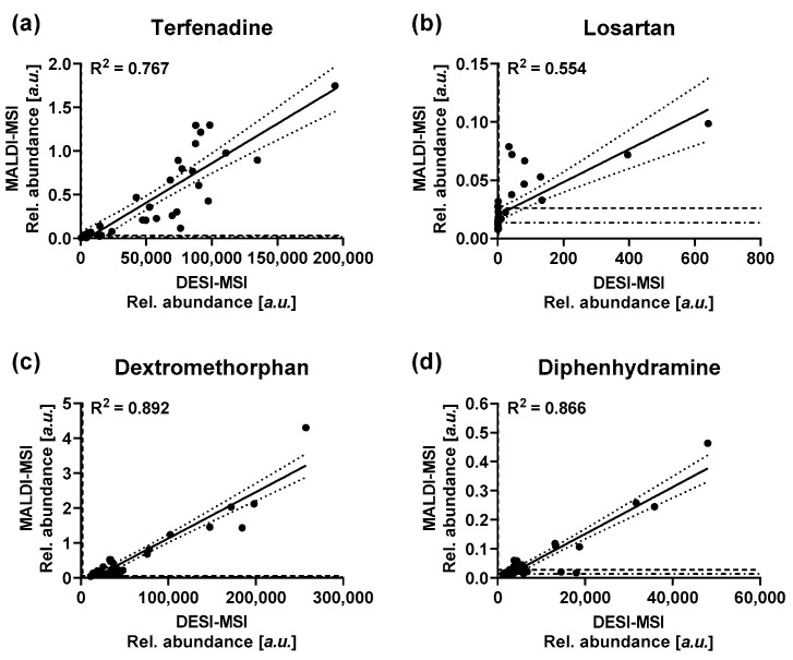
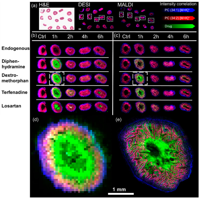
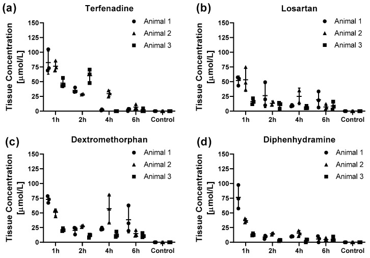
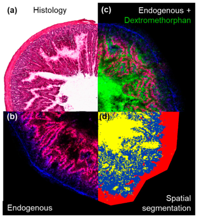
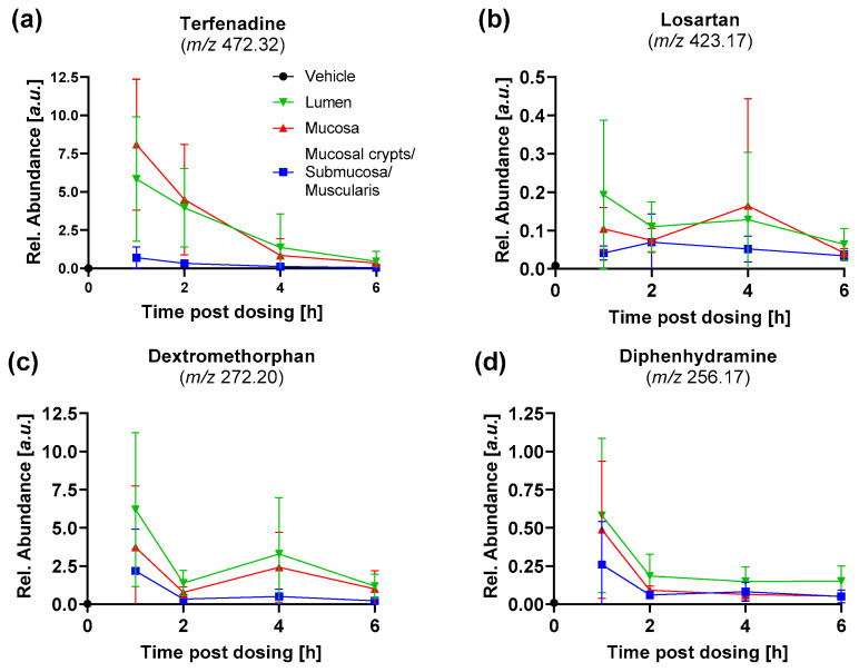
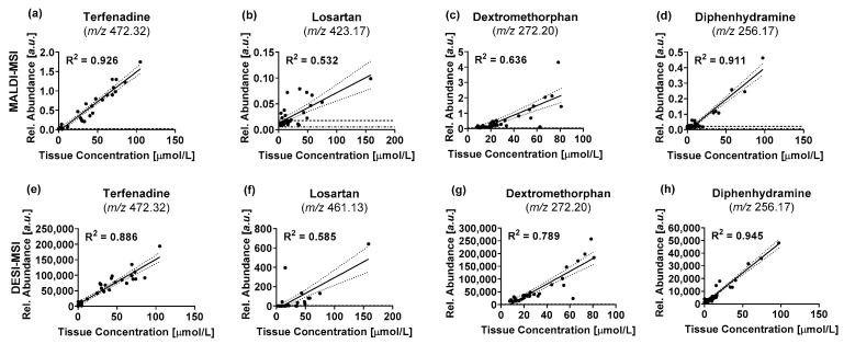
Liquid chromatography-tandem mass spectrometry (LC-MS/MS) is a standard tool used for absolute quantification of drugs in pharmacokinetic (PK) studies. However, all spatial information is lost during the extraction and elucidation of a drugs biodistribution within the tissue is impossible. In the study presented here we used a sample embedding protocol optimized for mass spectrometry imaging (MSI) to prepare up to 15 rat intestine specimens at once. Desorption electrospray ionization (DESI) and matrix assisted laser desorption/ionization (MALDI) mass spectrometry imaging (MSI) were employed to determine the distributions and relative abundances of four benchmarking compounds in the intestinal segments. High resolution MALDI-MSI experiments performed at 10 µm spatial resolution allowed to determine the drug distribution in the different intestinal histological compartments to determine the absorbed and tissue bound fractions of the drugs. The low tissue bound drug fractions, which were determined to account for 56-66% of the total drug, highlight the importance to understand the spatial distribution of drugs within the histological compartments of a given tissue to rationalize concentration differences found in PK studies. The mean drug abundances of four benchmark compounds determined by MSI were correlated with the absolute drug concentrations. Linear regression resulted in coefficients of determination (R) ranging from 0.532 to 0.926 for MALDI-MSI and R values ranging from 0.585 to 0.945 for DESI-MSI, validating a quantitative relation of the imaging data. The good correlation of the absolute tissue concentrations of the benchmark compounds and the MSI data provides a bases for relative quantification of compounds within and between tissues, without normalization to an isotopically labelled standard, provided that the compared tissues have inherently similar ion suppression effects.
液相色谱 - 串联质谱法(LC-MS/MS)是药代动力学(PK)研究中用于药物绝对定量的标准工具。然而,在提取过程中所有空间信息都会丢失,因此无法阐明药物在组织内的生物分布。在本文介绍的研究中,我们使用了一种针对质谱成像(MSI)优化的样本包埋方案,一次可制备多达15个大鼠肠道标本。采用解吸电喷雾电离(DESI)和基质辅助激光解吸/电离(MALDI)质谱成像(MSI)来确定四种基准化合物在肠道各段的分布和相对丰度。在10 µm空间分辨率下进行的高分辨率MALDI-MSI实验能够确定药物在不同肠道组织学区域的分布,以确定药物的吸收和组织结合部分。低组织结合药物部分占总药物的56 - 66%,这突出了了解药物在给定组织的组织学区域内的空间分布对于合理化PK研究中发现的浓度差异的重要性。通过MSI测定的四种基准化合物的平均药物丰度与绝对药物浓度相关。线性回归结果显示,MALDI-MSI的决定系数(R)范围为0.532至0.926,DESI-MSI的R值范围为0.585至0.945,验证了成像数据的定量关系。基准化合物的绝对组织浓度与MSI数据的良好相关性为组织内和组织间化合物的相对定量提供了基础,前提是所比较的组织具有本质上相似的离子抑制效应,无需对同位素标记标准进行归一化。