Gatin Eduard Gheorghe, Nagy Pal, Iordache Stefan-Marian, Iordache Ana-Maria, Luculescu Catalin Romeo
Faculty of Medicine, University of Medicine and Pharmacy "Carol Davila", 050474 Bucharest, Romania.
Faculty of Physics, University of Bucharest, 077125 Magurele, Romania.
Diagnostics (Basel). 2022 Mar 16;12(3):723. doi: 10.3390/diagnostics12030723.
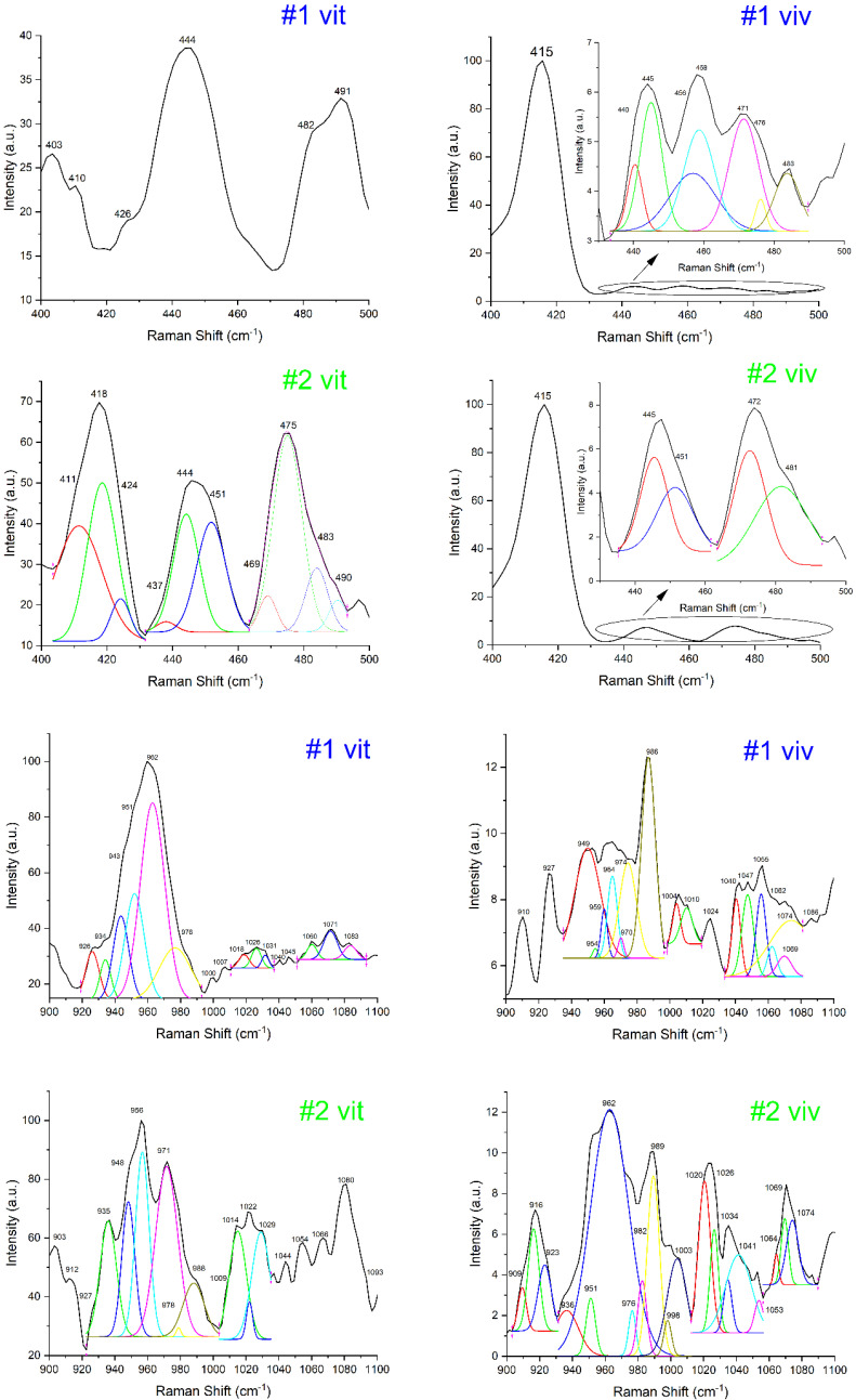
The aim of this study was to evaluate the quality of the bone, revealing the different phases for calcified tissues independent of the medical history of the patient in relation to periodontitis by means of in vivo Raman spectroscopy. Raman spectroscopy measurements were performed in vivo during surgery and then ex vivo for the harvested bone samples for the whole group of patients (ten patients). The specific peaks for the Raman spectrum were traced for reference compounds (e.g., calcium phosphates) and bone samples. The variation in the intensity of the spectrum in relation to the specific bone constituents' concentrations reflects the bone quality and can be strongly related with patient medical status (before dental surgery and after a healing period). Moreover, bone sample fluorescence is related to collagen content, enabling a complete evaluation of bone quality including a "quasi-quantification" of the healing process similar to the bone augmentation procedure. A complete evaluation of the processed spectra offers quantitative/qualitative information on the condition of the bone tissue. We conclude that Raman spectroscopy can be considered a viable investigation method for an in vivo and quick bone quality assessment during oral and periodontal surgery.
本研究的目的是通过体内拉曼光谱法评估骨质量,揭示钙化组织的不同阶段,且不考虑患者与牙周炎相关的病史。在手术过程中对整个患者组(10名患者)进行了体内拉曼光谱测量,然后对采集的骨样本进行了体外测量。针对参考化合物(如磷酸钙)和骨样本追踪拉曼光谱的特定峰。光谱强度相对于特定骨成分浓度的变化反映了骨质量,并且可能与患者的医疗状况(牙科手术前和愈合期后)密切相关。此外,骨样本荧光与胶原蛋白含量相关,能够对骨质量进行全面评估,包括对类似于骨增量手术的愈合过程进行“准定量”。对处理后的光谱进行全面评估可提供有关骨组织状况的定量/定性信息。我们得出结论,拉曼光谱可被视为一种可行的研究方法,用于在口腔和牙周手术期间进行体内快速骨质量评估。