• 文献检索
  • 文档翻译
  • 深度研究
  • 学术资讯
  • Suppr Zotero 插件Zotero 插件
  • 邀请有礼
  • 套餐&价格
  • 历史记录
应用&插件
Suppr Zotero 插件Zotero 插件浏览器插件Mac 客户端Windows 客户端微信小程序
定价
高级版会员购买积分包购买API积分包
服务
文献检索文档翻译深度研究API 文档MCP 服务
关于我们
关于 Suppr公司介绍联系我们用户协议隐私条款
关注我们

Suppr 超能文献

核心技术专利:CN118964589B侵权必究
粤ICP备2023148730 号-1Suppr @ 2026

文献检索

告别复杂PubMed语法,用中文像聊天一样搜索,搜遍4000万医学文献。AI智能推荐,让科研检索更轻松。

立即免费搜索

文件翻译

保留排版,准确专业,支持PDF/Word/PPT等文件格式,支持 12+语言互译。

免费翻译文档

深度研究

AI帮你快速写综述,25分钟生成高质量综述,智能提取关键信息,辅助科研写作。

立即免费体验

非流行地区基于视觉评估和人工智能评估的CO-RADS评分比较

Comparison of CO-RADS Scores Based on Visual and Artificial Intelligence Assessments in a Non-Endemic Area.

作者信息

Ishiwata Yoshinobu, Miura Kentaro, Kishimoto Mayuko, Nomura Koichiro, Sawamura Shungo, Magami Shigeru, Ikawa Mizuki, Yamashiro Tsuneo, Utsunomiya Daisuke

机构信息

Department of Radiology, Yokohama City University Hospital, 3-9 Fukuura, Kanazawa-ku, Yokohama 236-0004, Japan.

Department of Radiology, Yokohama Municipal Citizen's Hospital, 1-1 Mitsuzawa Nishimachi, Kanagawa-ku, Yokohama 221-0855, Japan.

出版信息

Diagnostics (Basel). 2022 Mar 18;12(3):738. doi: 10.3390/diagnostics12030738.

DOI:10.3390/diagnostics12030738
PMID:35328290
原文链接:https://pmc.ncbi.nlm.nih.gov/articles/PMC8946998/
Abstract

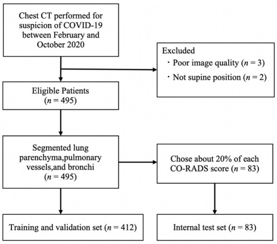
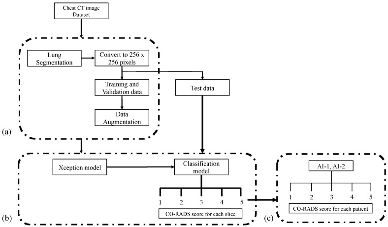
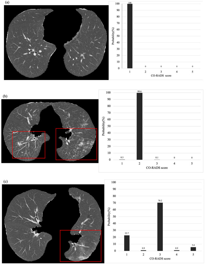
In this study, we first developed an artificial intelligence (AI)-based algorithm for classifying chest computed tomography (CT) images using the coronavirus disease 2019 Reporting and Data System (CO-RADS). Subsequently, we evaluated its accuracy by comparing the calculated scores with those assigned by radiologists with varying levels of experience. This study included patients with suspected SARS-CoV-2 infection who underwent chest CT imaging between February and October 2020 in Japan, a non-endemic area. For each chest CT, the CO-RADS scores, determined by consensus among three experienced chest radiologists, were used as the gold standard. Images from 412 patients were used to train the model, whereas images from 83 patients were tested to obtain AI-based CO-RADS scores for each image. Six independent raters (one medical student, two residents, and three board-certified radiologists) evaluated the test images. Intraclass correlation coefficients (ICC) and weighted kappa values were calculated to determine the inter-rater agreement with the gold standard. The mean ICC and weighted kappa were 0.754 and 0.752 for the medical student and residents (taken together), 0.851 and 0.850 for the diagnostic radiologists, and 0.913 and 0.912 for AI, respectively. The CO-RADS scores calculated using our AI-based algorithm were comparable to those assigned by radiologists, indicating the accuracy and high reproducibility of our model. Our study findings would enable accurate reading, particularly in areas where radiologists are unavailable, and contribute to improvements in patient management and workflow.

摘要

在本研究中,我们首先开发了一种基于人工智能(AI)的算法,用于使用2019冠状病毒病报告和数据系统(CO-RADS)对胸部计算机断层扫描(CT)图像进行分类。随后,我们通过将计算出的分数与不同经验水平的放射科医生给出的分数进行比较,评估了其准确性。本研究纳入了2020年2月至10月在日本(一个非流行地区)接受胸部CT成像的疑似SARS-CoV-2感染患者。对于每例胸部CT,由三位经验丰富的胸部放射科医生通过共识确定的CO-RADS分数用作金标准。来自412例患者的图像用于训练模型,而来自83例患者的图像用于测试,以获得每张图像基于AI的CO-RADS分数。六位独立评估者(一名医学生、两名住院医师和三名获得委员会认证的放射科医生)对测试图像进行了评估。计算组内相关系数(ICC)和加权kappa值,以确定评估者与金标准之间的一致性。医学生和住院医师(合在一起)的平均ICC和加权kappa分别为0.754和0.752,诊断放射科医生为0.851和0.850,AI为0.913和0.912。使用我们基于AI的算法计算出的CO-RADS分数与放射科医生给出的分数相当,表明我们模型的准确性和高重复性。我们的研究结果将有助于进行准确的解读,特别是在没有放射科医生的地区,并有助于改善患者管理和工作流程。

https://cdn.ncbi.nlm.nih.gov/pmc/blobs/75a7/8946998/efa8a896661c/diagnostics-12-00738-g005.jpg
https://cdn.ncbi.nlm.nih.gov/pmc/blobs/75a7/8946998/3c9fde2a91ae/diagnostics-12-00738-g001.jpg
https://cdn.ncbi.nlm.nih.gov/pmc/blobs/75a7/8946998/5f4fb6d8f84b/diagnostics-12-00738-g002.jpg
https://cdn.ncbi.nlm.nih.gov/pmc/blobs/75a7/8946998/b3313e573b9f/diagnostics-12-00738-g003.jpg
https://cdn.ncbi.nlm.nih.gov/pmc/blobs/75a7/8946998/e8a758762b1f/diagnostics-12-00738-g004a.jpg
https://cdn.ncbi.nlm.nih.gov/pmc/blobs/75a7/8946998/efa8a896661c/diagnostics-12-00738-g005.jpg

相似文献

1
Comparison of CO-RADS Scores Based on Visual and Artificial Intelligence Assessments in a Non-Endemic Area.非流行地区基于视觉评估和人工智能评估的CO-RADS评分比较
Diagnostics (Basel). 2022 Mar 18;12(3):738. doi: 10.3390/diagnostics12030738.
2
Automated Assessment of COVID-19 Reporting and Data System and Chest CT Severity Scores in Patients Suspected of Having COVID-19 Using Artificial Intelligence.利用人工智能对疑似 COVID-19 患者的 COVID-19 报告和数据系统及胸部 CT 严重程度评分进行自动评估。
Radiology. 2021 Jan;298(1):E18-E28. doi: 10.1148/radiol.2020202439. Epub 2020 Jul 30.
3
Radiological Society of North America (RSNA) Expert Consensus Statement Related to Chest CT Findings in COVID-19 Versus CO-RADS: Comparison of Reporting System Performance Among Chest Radiologists and End-User Preference.北美放射学会(RSNA)关于 COVID-19 与 CO-RADS 胸部 CT 表现的专家共识声明:胸部放射科医生和最终用户偏好之间报告系统性能的比较。
Can Assoc Radiol J. 2021 Nov;72(4):806-813. doi: 10.1177/0846537120968919. Epub 2020 Nov 3.
4
Diagnostic accuracy and inter-observer agreement with the CO-RADS lexicon for CT chest reporting in COVID-19.CO-RADS 词汇在 COVID-19 胸部 CT 报告中的诊断准确性和观察者间一致性。
Emerg Radiol. 2021 Dec;28(6):1045-1054. doi: 10.1007/s10140-021-01967-6. Epub 2021 Jul 24.
5
Artificial Intelligence Algorithm Detecting Lung Infection in Supine Chest Radiographs of Critically Ill Patients With a Diagnostic Accuracy Similar to Board-Certified Radiologists.人工智能算法检测重症患者仰卧位胸部 X 光片肺部感染的诊断准确性与放射科认证医师相当。
Crit Care Med. 2020 Jul;48(7):e574-e583. doi: 10.1097/CCM.0000000000004397.
6
Is the generalizability of a developed artificial intelligence algorithm for COVID-19 on chest CT sufficient for clinical use? Results from the International Consortium for COVID-19 Imaging AI (ICOVAI).开发用于 COVID-19 胸部 CT 的人工智能算法的可推广性是否足以用于临床?来自 COVID-19 成像人工智能国际联合会(ICOVAI)的结果。
Eur Radiol. 2023 Jun;33(6):4249-4258. doi: 10.1007/s00330-022-09303-3. Epub 2023 Jan 18.
7
Differing benefits of artificial intelligence-based computer-aided diagnosis for breast US according to workflow and experience level.根据工作流程和经验水平,基于人工智能的乳腺超声计算机辅助诊断具有不同的益处。
Ultrasonography. 2022 Oct;41(4):718-727. doi: 10.14366/usg.22014. Epub 2022 Mar 30.
8
Comparison of Chest Radiograph Interpretations by Artificial Intelligence Algorithm vs Radiology Residents.人工智能算法与放射科住院医师对胸部 X 线片解读的比较。
JAMA Netw Open. 2020 Oct 1;3(10):e2022779. doi: 10.1001/jamanetworkopen.2020.22779.
9
Combination of Deep Learning-Based Denoising and Iterative Reconstruction for Ultra-Low-Dose CT of the Chest: Image Quality and Lung-RADS Evaluation.基于深度学习的降噪与迭代重建在胸部超低剂量 CT 中的联合应用:图像质量与 Lung-RADS 评估。
AJR Am J Roentgenol. 2020 Dec;215(6):1321-1328. doi: 10.2214/AJR.19.22680. Epub 2020 Oct 14.
10
Evaluation of the Usefulness of CO-RADS for Chest CT in Patients Suspected of Having COVID-19.CO-RADS在疑似新型冠状病毒肺炎患者胸部CT中的应用价值评估
Diagnostics (Basel). 2020 Aug 19;10(9):608. doi: 10.3390/diagnostics10090608.

本文引用的文献

1
Why Does the SARS-CoV-2 Delta VOC Spread So Rapidly? Universal Conditions for the Rapid Spread of Respiratory Viruses, Minimum Viral Loads for Viral Aerosol Generation, Effects of Vaccination on Viral Aerosol Generation, and Viral Aerosol Clouds.为什么 SARS-CoV-2 德尔塔 VOC 传播如此迅速?呼吸道病毒快速传播的普遍条件、病毒气溶胶产生的最小病毒载量、疫苗接种对病毒气溶胶产生的影响以及病毒气溶胶云。
Int J Environ Res Public Health. 2021 Sep 17;18(18):9804. doi: 10.3390/ijerph18189804.
2
Real-world evaluation of a computed tomography-first triage strategy for suspected Coronavirus disease 2019 in outpatients in Japan: An observational cohort study.日本门诊疑似 2019 冠状病毒病患者行计算机断层扫描优先分诊策略的真实世界评价:一项观察性队列研究。
Medicine (Baltimore). 2021 Jun 4;100(22):e26161. doi: 10.1097/MD.0000000000026161.
3
A meta-analysis of accuracy and sensitivity of chest CT and RT-PCR in COVID-19 diagnosis.一项关于 COVID-19 诊断中胸部 CT 和 RT-PCR 准确性和敏感性的荟萃分析。
Sci Rep. 2020 Dec 28;10(1):22402. doi: 10.1038/s41598-020-80061-2.
4
A head-to-head comparison of the intra- and interobserver agreement of COVID-RADS and CO-RADS grading systems in a population with high estimated prevalence of COVID-19.在新冠肺炎估计患病率较高的人群中,对COVID-RADS和CO-RADS分级系统的观察者内和观察者间一致性进行的直接比较。
BJR Open. 2020 Dec 11;2(1):20200053. doi: 10.1259/bjro.20200053. eCollection 2020.
5
Chest CT in COVID-19 at the ED: Validation of the COVID-19 Reporting and Data System (CO-RADS) and CT Severity Score: A Prospective, Multicenter, Observational Study.急诊科 COVID-19 的胸部 CT:COVID-19 报告和数据系统 (CO-RADS) 和 CT 严重程度评分的验证:一项前瞻性、多中心、观察性研究。
Chest. 2021 Mar;159(3):1126-1135. doi: 10.1016/j.chest.2020.11.026. Epub 2020 Nov 30.
6
Visualizing "featureless" regions on mammograms classified as invasive ductal carcinomas by a deep learning algorithm: the promise of AI support in radiology.利用深度学习算法对浸润性导管癌进行分类的乳腺 X 光片中“无特征”区域的可视化:人工智能在放射学中的应用前景。
Jpn J Radiol. 2021 Apr;39(4):333-340. doi: 10.1007/s11604-020-01070-9. Epub 2020 Nov 16.
7
Early prediction of disease progression in COVID-19 pneumonia patients with chest CT and clinical characteristics.基于胸部 CT 与临床特征的 COVID-19 肺炎患者疾病进展的早期预测。
Nat Commun. 2020 Oct 2;11(1):4968. doi: 10.1038/s41467-020-18786-x.
8
Diagnostic accuracy and interobserver variability of CO-RADS in patients with suspected coronavirus disease-2019: a multireader validation study.CO-RADS 在疑似 2019 冠状病毒病患者中的诊断准确性和观察者间变异性:一项多读者验证研究。
Eur Radiol. 2021 Apr;31(4):1932-1940. doi: 10.1007/s00330-020-07273-y. Epub 2020 Sep 23.
9
Predictors of COVID-19 severity: A literature review.预测 COVID-19 严重程度的因素:文献综述。
Rev Med Virol. 2021 Jan;31(1):1-10. doi: 10.1002/rmv.2146. Epub 2020 Jul 30.
10
Evaluation of the Usefulness of CO-RADS for Chest CT in Patients Suspected of Having COVID-19.CO-RADS在疑似新型冠状病毒肺炎患者胸部CT中的应用价值评估
Diagnostics (Basel). 2020 Aug 19;10(9):608. doi: 10.3390/diagnostics10090608.