Institute of Neuropathology, RWTH Aachen University Hospital, 52074 Aachen, Germany.
Department of Neurosurgery, RWTH Aachen University Hospital, 52074 Aachen, Germany.
Int J Mol Sci. 2022 Mar 16;23(6):3221. doi: 10.3390/ijms23063221.
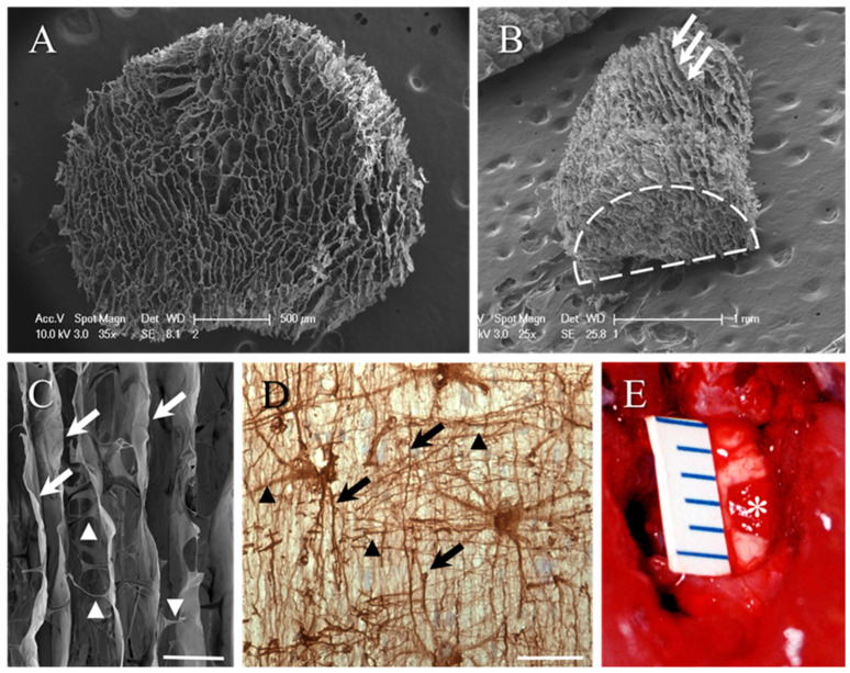
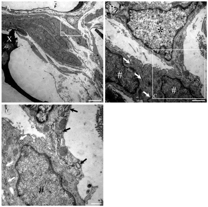
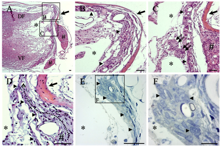
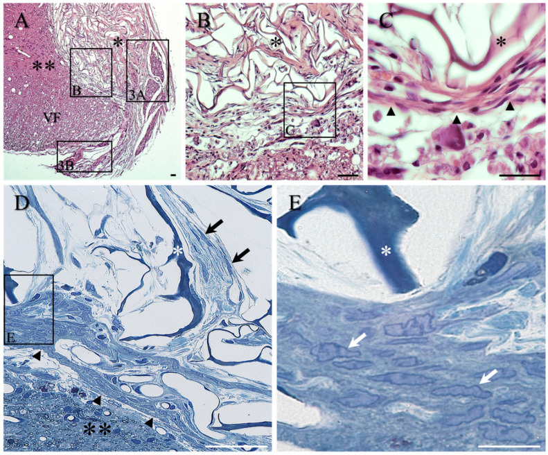
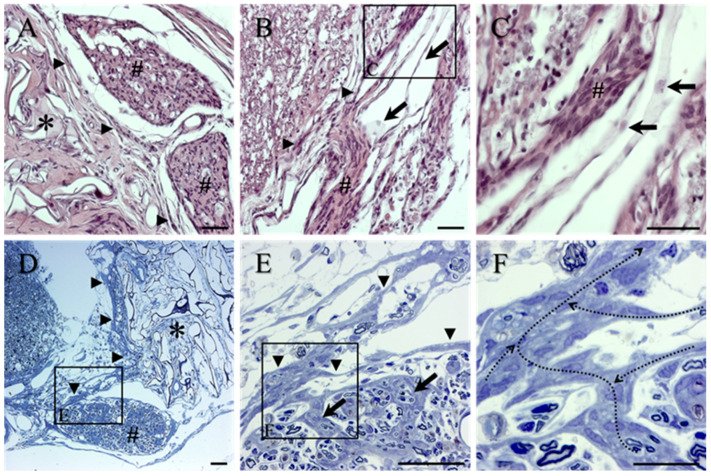
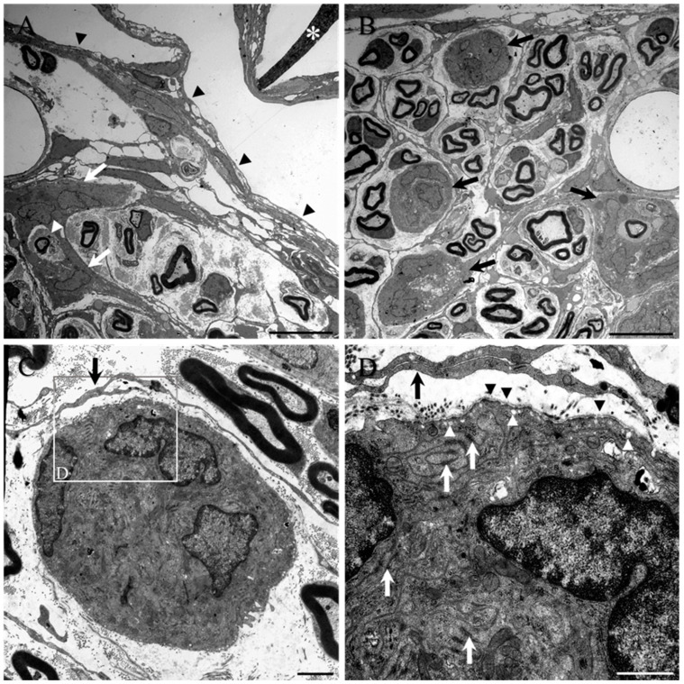
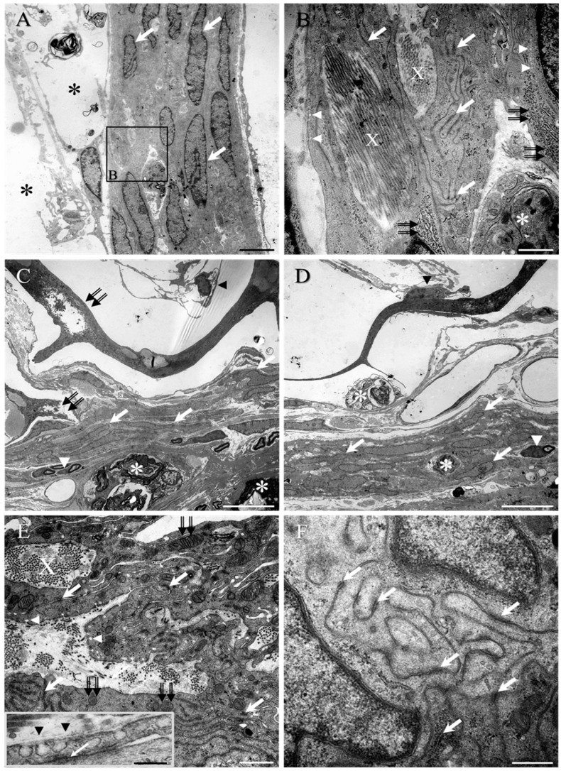
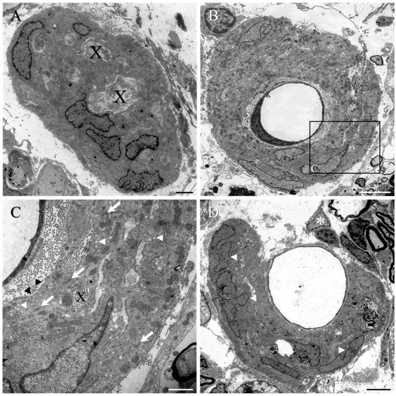
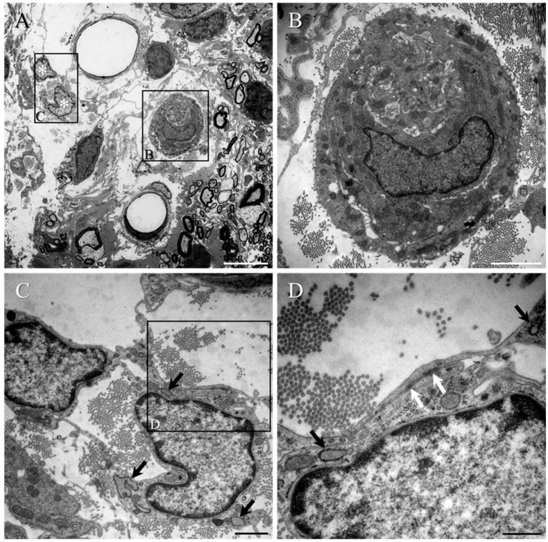
Numerous intervention strategies have been developed to promote functional tissue repair following experimental spinal cord injury (SCI), including the bridging of lesion-induced cystic cavities with bioengineered scaffolds. Integration between such implanted scaffolds and the lesioned host spinal cord is critical for supporting regenerative growth, but only moderate-to-low degrees of success have been reported. Light and electron microscopy were employed to better characterise the fibroadhesive scarring process taking place after implantation of a longitudinally microstructured type-I collagen scaffold into unilateral mid-cervical resection injuries of the adult rat spinal cord. At long survival times (10 weeks post-surgery), sheets of tightly packed cells (of uniform morphology) could be seen lining the inner surface of the repaired dura mater of lesion-only control animals, as well as forming a barrier along the implant-host interface of the scaffold-implanted animals. The highly uniform ultrastructural features of these scarring cells and their anatomical continuity with the local, reactive spinal nerve roots strongly suggest their identity to be perineurial-like cells. This novel aspect of the cellular composition of reactive spinal cord tissue highlights the increasingly complex nature of fibroadhesive scarring involved in traumatic injury, and particularly in response to the implantation of bioengineered collagen scaffolds.
已经开发出许多干预策略来促进实验性脊髓损伤 (SCI) 后的功能性组织修复,包括用生物工程支架桥接损伤引起的囊性腔。这些植入支架与损伤宿主脊髓之间的整合对于支持再生性生长至关重要,但据报道,只有中等至低程度的成功。采用光学显微镜和电子显微镜更好地描述了将纵向微结构化 I 型胶原支架植入成年大鼠脊髓单侧颈中部切除损伤后的纤维粘连性瘢痕形成过程。在长期存活时间(手术后 10 周),仅损伤对照动物的修复硬脑膜内表面可以看到紧密堆积的细胞(形态均匀)的薄片,以及在支架植入动物的植入物-宿主界面沿线形成屏障。这些瘢痕细胞的高度均匀的超微结构特征及其与局部反应性脊神经根的解剖连续性强烈表明其为许旺氏细胞样细胞。反应性脊髓组织细胞成分的这一新方面突出了外伤性损伤中纤维粘连性瘢痕形成的日益复杂性质,特别是对生物工程胶原支架植入的反应。