Regulski Piotr A, Zielinski Jakub, Szopinski Kazimierz T
Department of Dental and Maxillofacial Radiology, Faculty of Medicine and Dentistry, Medical University of Warsaw, 61 Zwirki i Wigury Street, 02-091 Warsaw, Poland.
Interdisciplinary Centre for Mathematical and Computational Modelling, University of Warsaw, Krakowskie Przedmiescie 26/28, 00-927 Warsaw, Poland.
J Clin Med. 2022 Mar 15;11(6):1621. doi: 10.3390/jcm11061621.
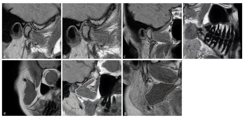
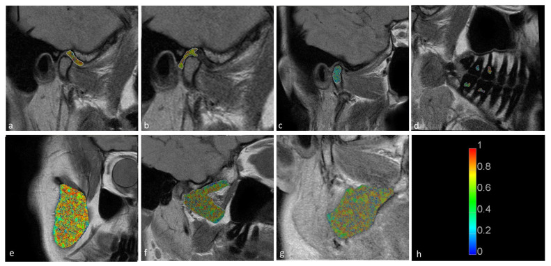
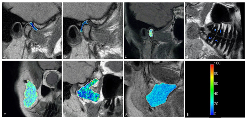
In this study, we aimed to assess the potential impact of temporomandibular disk displacement on anatomical structures of the stomatognathic system using biexponential T2 magnetic resonance imaging (MRI) maps. Fifty separate MRI scans of the temporomandibular joints (TMJ) of 25 patients were acquired with eight echo times. Biexponential T2 maps were created by weighted reconstruction based on Powell's conjugate direction method and divided into two groups: the TMJ without (32 images) and with (18 images) disk displacement. The disk, retrodiscal tissue, condylar bone marrow, masseter muscle, lateral and medial pterygoid muscles and dental pulp of the first and second molars were manually segmented twice. The intrarater reliability was assessed. The averages and standard deviations of the T2 times and fractions of each segmented region for each group were calculated and analysed with multiple Student's -tests. Significant differences between groups were observed in the retrodiscal tissue, medial pterygoid muscle and bone marrow. The pulp short T2 component showed a trend toward statistical significance. The segmentation reliability was excellent (93.6%). The relationship between disk displacement and quantitative MRI features of stomatognathic structures can be useful in the combined treatment of articular disk displacement, pterygoid muscle tension and occlusive reconstruction.
在本研究中,我们旨在使用双指数T2磁共振成像(MRI)图谱评估颞下颌关节盘移位对口腔颌面部系统解剖结构的潜在影响。对25例患者的颞下颌关节(TMJ)进行了50次独立的MRI扫描,采集了8个回波时间的数据。基于鲍威尔共轭方向法通过加权重建创建双指数T2图谱,并将其分为两组:无关节盘移位的颞下颌关节(32幅图像)和有关节盘移位的颞下颌关节(18幅图像)。对关节盘、盘后组织、髁突骨髓、咬肌、翼外肌和翼内肌以及第一和第二磨牙的牙髓进行了两次手动分割。评估了评分者内信度。计算每组每个分割区域的T2时间和分数的平均值和标准差,并进行多次学生t检验分析。在盘后组织、翼内肌和骨髓中观察到组间存在显著差异。牙髓短T2成分显示出具有统计学意义的趋势。分割可靠性极佳(93.6%)。关节盘移位与口腔颌面部结构的定量MRI特征之间的关系在关节盘移位、翼肌紧张和咬合重建的联合治疗中可能有用。