• 文献检索
  • 文档翻译
  • 深度研究
  • 学术资讯
  • Suppr Zotero 插件Zotero 插件
  • 邀请有礼
  • 套餐&价格
  • 历史记录
应用&插件
Suppr Zotero 插件Zotero 插件浏览器插件Mac 客户端Windows 客户端微信小程序
定价
高级版会员购买积分包购买API积分包
服务
文献检索文档翻译深度研究API 文档MCP 服务
关于我们
关于 Suppr公司介绍联系我们用户协议隐私条款
关注我们

Suppr 超能文献

核心技术专利:CN118964589B侵权必究
粤ICP备2023148730 号-1Suppr @ 2026

文献检索

告别复杂PubMed语法,用中文像聊天一样搜索,搜遍4000万医学文献。AI智能推荐,让科研检索更轻松。

立即免费搜索

文件翻译

保留排版,准确专业,支持PDF/Word/PPT等文件格式,支持 12+语言互译。

免费翻译文档

深度研究

AI帮你快速写综述,25分钟生成高质量综述,智能提取关键信息,辅助科研写作。

立即免费体验

颞下颌关节(TMJ)疼痛患者的临床检查结果与磁共振成像(MRI)结果是否相关?一项横断面研究。

Does clinical findings correlate with magnetic resonance imaging (MRI) findings in patients with temporomandibular joint (TMJ) pain? A cross sectional study.

机构信息

Department of Oral and Maxillofacial Surgery Faculty of Dentistry, Recep Tayyip Erdoğan University Rize, Turkey

出版信息

Med Oral Patol Oral Cir Bucal. 2020 Jul 1;25(4):e495-e501. doi: 10.4317/medoral.23501.

DOI:10.4317/medoral.23501
PMID:32134894
原文链接:https://pmc.ncbi.nlm.nih.gov/articles/PMC7338075/
Abstract

BACKGROUND

Although magnetic resonance imaging (MRI) helps to clearly visualize the disorders in temporomandibular joint (TMJ), the relationship between cross-sectional and clinical findings has not been precisely established. The aim of this study was to evaluate the relationship between clinical symptoms and MRI findings in individuals with TMJ pain.

MATERIAL AND METHODS

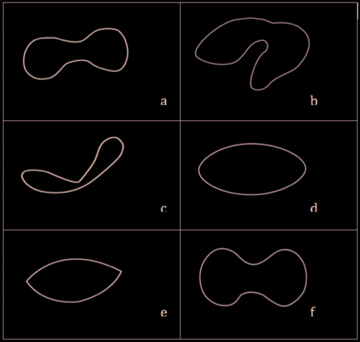
This cross-sectional study, conducted on the clinical and MRI findings of the patients, who applied to Uşak University, Oral and Maxillofacial Surgery Clinic with TMJ pain between the years 2016-2019. The primary predictor variables were MRI findings; disc position (normal, disc displacement with reduction (DDWR), disc displacement without reduction (DDWOR)), disc structural distortion (normal, folded, lengthened, round, biconvex, thick), condyle degeneration type (normal, moderate, severe) and joint effusion (JE) (absent, present). The primary outcome variable was pain, recorded on a visual analog scale (VAS) (numbered between 0-10). The other variables were demographic variables (age/gender). The relationship between clinical and MRI findings were statistically evaluated. The data were analysed by Kruskal Wallis and Mann Whitney U test. Chi-square (x2) test was used for categorical variable comparisons. P values < .05 were considered to indicate statistical significance.

RESULTS

Clinical and MRI records of 700 TMJ, from 350 patients with the mean age of the 31 (12-65) were evaluated in this study. Statistically significant differences were found between; disc position and pain, disc position and JE; JE and pain; disc structural distortion and pain; and disc structural distortion and disc position. JE was seen more common in DDWOR group. The most common disc distortion, seen in patients with JE, is the folded type.

CONCLUSIONS

The present study can infer that pain is associated with disc position, JE, disc structural distortion, and DDWOR is associated with JE. Folded type disc is the most common disc type in TMJ with JE.

摘要

背景

尽管磁共振成像(MRI)有助于清晰地观察颞下颌关节(TMJ)的病变,但横断面和临床发现之间的关系尚未精确确定。本研究旨在评估 TMJ 疼痛患者的临床症状与 MRI 发现之间的关系。

材料和方法

这是一项横断面研究,对 2016 年至 2019 年期间因 TMJ 疼痛到乌沙克大学口腔颌面外科诊所就诊的患者的临床和 MRI 检查结果进行了研究。主要预测变量是 MRI 结果;盘位置(正常、盘移位伴复位(DDWR)、盘移位无复位(DDWOR))、盘结构变形(正常、折叠、延长、圆形、双凸、增厚)、髁突退变类型(正常、中度、重度)和关节积液(JE)(无、有)。主要结局变量是疼痛,用视觉模拟评分(VAS)记录(编号为 0-10)。其他变量为人口统计学变量(年龄/性别)。对临床和 MRI 结果之间的关系进行了统计学评估。数据通过 Kruskal Wallis 和 Mann Whitney U 检验进行分析。卡方(x2)检验用于分类变量比较。P 值<.05 表示具有统计学意义。

结果

本研究共评估了 350 名患者的 700 个 TMJ 的临床和 MRI 记录,平均年龄为 31 岁(12-65 岁)。盘位置与疼痛、盘位置与 JE、JE 与疼痛、盘结构变形与疼痛以及盘结构变形与盘位置之间存在统计学显著差异。DDWOR 组中 JE 更为常见。在有 JE 的患者中,最常见的盘变形是折叠型。

结论

本研究可以推断出疼痛与盘位置、JE、盘结构变形有关,DDWOR 与 JE 有关。在有 JE 的 TMJ 中,最常见的盘类型是折叠型。

https://cdn.ncbi.nlm.nih.gov/pmc/blobs/a64f/7338075/061a7c8e2c8f/medoral-25-e495-g001.jpg

相似文献

1
Does clinical findings correlate with magnetic resonance imaging (MRI) findings in patients with temporomandibular joint (TMJ) pain? A cross sectional study.颞下颌关节(TMJ)疼痛患者的临床检查结果与磁共振成像(MRI)结果是否相关?一项横断面研究。
Med Oral Patol Oral Cir Bucal. 2020 Jul 1;25(4):e495-e501. doi: 10.4317/medoral.23501.
2
Effects of bruxism on temporomandibular joint internal derangement in patients with unilateral temporomandibular joint pain: The role of magnetic resonance imaging diagnostics.磨牙症对单侧颞下颌关节疼痛患者颞下颌关节内紊乱的影响:磁共振成像诊断的作用
Cranio. 2024 Mar;42(2):113-121. doi: 10.1080/08869634.2021.1918959. Epub 2021 Apr 25.
3
Temporomandibular joint internal derangement: relationship between joint pain and MR grading of effusion and total protein concentration in the joint fluid.颞下颌关节内紊乱:关节疼痛与关节积液的磁共振成像分级及关节液总蛋白浓度之间的关系
Dentomaxillofac Radiol. 2005 May;34(3):175-81. doi: 10.1259/dmfr/49181266.
4
[MRI observation of condylar location and morphology in the patients with temporomandibular disc displacement].[颞下颌关节盘移位患者髁突位置及形态的MRI观察]
Zhonghua Kou Qiang Yi Xue Za Zhi. 2019 Aug 9;54(8):522-526. doi: 10.3760/cma.j.issn.1002-0098.2019.08.004.
5
Association between temporomandibular joint disc position evaluated by magnetic resonance imaging and mandibular condyle inclination evaluated by computed tomography.磁共振成像评估的颞下颌关节盘位置与计算机断层扫描评估的髁突倾斜度之间的关系。
J Oral Rehabil. 2020 Jun;47(6):743-749. doi: 10.1111/joor.12962. Epub 2020 Apr 24.
6
MRI investigation of TMJ disc and articular eminence morphology in patients with disc displacement.MRI 对颞下颌关节盘和关节突形态的研究在盘移位患者中的应用。
J Stomatol Oral Maxillofac Surg. 2021 Feb;122(1):3-6. doi: 10.1016/j.jormas.2020.09.019. Epub 2020 Oct 12.
7
Ultrasound assessment of increased capsular width in temporomandibular joint internal derangements: relationship with joint pain and magnetic resonance grading of joint effusion.颞下颌关节内紊乱中关节囊宽度增加的超声评估:与关节疼痛及关节积液磁共振分级的关系
Oral Surg Oral Med Oral Pathol Oral Radiol Endod. 2011 Jul;112(1):112-7. doi: 10.1016/j.tripleo.2011.02.020. Epub 2011 May 4.
8
Does Temporomandibular Joint Magnetic Resonance Imaging Diagnosis Support Clinical Examination Diagnosis Following Diagnostic Criteria for Temporomandibular Disorders?颞下颌关节磁共振成像诊断是否支持根据颞下颌关节紊乱病的诊断标准进行临床检查诊断?
J Oral Maxillofac Surg. 2023 Jul;81(7):813-819. doi: 10.1016/j.joms.2023.03.007. Epub 2023 Mar 28.
9
Comparison of imaging findings of 714 symptomatic and asymptomatic temporomandibular joints: a retrospective study.714 例有症状和无症状颞下颌关节影像学表现的对比:一项回顾性研究。
BMC Oral Health. 2023 Feb 7;23(1):79. doi: 10.1186/s12903-023-02783-9.
10
Temporomandibular joint disorders' impact on pain, function, and disability.颞下颌关节紊乱症对疼痛、功能和残疾的影响。
J Dent Res. 2015 Mar;94(3 Suppl):79S-86S. doi: 10.1177/0022034514565793. Epub 2015 Jan 8.

引用本文的文献

1
The influence of open disc repositioning surgery on the internal derangement of the contralateral temporomandibular joint: a prospective study of 96 patients.开口复位手术对健侧颞下颌关节内部紊乱的影响:96 例患者的前瞻性研究。
Clin Oral Investig. 2024 Aug 15;28(9):487. doi: 10.1007/s00784-024-05864-3.
2
Comparative study of clinical and MRI features of TMD patients with or without joint effusion: a retrospective study.伴或不伴关节积液的 TMD 患者的临床和 MRI 特征的对比研究:一项回顾性研究。
BMC Oral Health. 2024 Mar 9;24(1):314. doi: 10.1186/s12903-024-04065-4.
3
Temporomandibular Disk Dislocation Impacts the Stomatognathic System: Comparative Study Based on Biexponential Quantitative T2 Maps.

本文引用的文献

1
Evaluation of serum prolidase activity and oxidative stress in patients with temporomandibular joint internal derangement.评价颞下颌关节紊乱症患者血清脯氨酸酶活性和氧化应激水平。
Cranio. 2021 May;39(3):238-248. doi: 10.1080/08869634.2019.1606987. Epub 2019 Apr 25.
2
The relationship between temporomandibular joint effusion and pain in patients with internal derangement.关节积液与颞下颌关节紊乱病患者疼痛的关系。
J Craniomaxillofac Surg. 2019 Jun;47(6):940-944. doi: 10.1016/j.jcms.2019.03.010. Epub 2019 Mar 15.
3
Correlation between joint effusion and morphology of the articular disc within the temporomandibular joint as viewed in the sagittal plane in patients with chronic disc displacement with reduction: A retrospective analytical study from magnetic resonance imaging.
颞下颌关节盘脱位对口腔颌面部系统的影响:基于双指数定量T2图的比较研究
J Clin Med. 2022 Mar 15;11(6):1621. doi: 10.3390/jcm11061621.
4
Analysis of three-dimensional imaging findings and clinical symptoms in patients with temporomandibular joint disorders.颞下颌关节紊乱病患者的三维成像结果与临床症状分析
Quant Imaging Med Surg. 2021 May;11(5):1921-1931. doi: 10.21037/qims-20-857.
慢性可复性盘前移位患者关节盘矢状面 MRI 关节腔积液与关节盘形态的相关性:回顾性分析研究。
Cranio. 2021 Mar;39(2):119-124. doi: 10.1080/08869634.2019.1582166. Epub 2019 Feb 21.
4
Patient symptoms and magnetic resonance imaging correlation in temporomandibular joint internal derangement.颞下颌关节内紊乱的患者症状与磁共振成像的相关性
Turk J Med Sci. 2018 Dec 12;48(6):1092-1095. doi: 10.3906/sag-1707-173.
5
Association of Temporomandibular Joint Pain According to Magnetic Resonance Imaging Findings in Temporomandibular Disorder Patients.颞下颌关节紊乱病患者磁共振成像结果与颞下颌关节疼痛的相关性
J Oral Maxillofac Surg. 2017 Sep;75(9):1848-1855. doi: 10.1016/j.joms.2017.03.026. Epub 2017 Mar 24.
6
The value of MRI in patients with temporomandibular joint dysfunction: Correlation of MRI and clinical findings.磁共振成像在颞下颌关节紊乱病患者中的价值:磁共振成像与临床检查结果的相关性
Eur J Radiol. 2016 Apr;85(4):714-9. doi: 10.1016/j.ejrad.2016.02.001. Epub 2016 Feb 4.
7
Correlation Between Clinical Findings of Temporomandibular Disorders and MRI Characteristics of Disc Displacement.颞下颌关节紊乱病临床症状与盘移位MRI特征的相关性
Open Dent J. 2015 Jul 31;9:273-81. doi: 10.2174/1874210601509010273. eCollection 2015.
8
Relationship between anterior disc displacement with/without reduction and effusion in temporomandibular disorder patients using magnetic resonance imaging.使用磁共振成像研究颞下颌关节紊乱症患者中伴或不伴可复性的关节盘前移位与关节积液之间的关系。
Imaging Sci Dent. 2013 Dec;43(4):245-51. doi: 10.5624/isd.2013.43.4.245. Epub 2013 Dec 12.
9
Prevalence of temporomandibular joint disorders among students of the university of jordan.约旦大学学生颞下颌关节紊乱症的患病率
J Clin Med Res. 2009 Aug;1(3):158-64. doi: 10.4021/jocmr2009.06.1245. Epub 2009 Jul 3.
10
MRI analysis of the relationship between bone changes in the temporomandibular joint and articular disc position in symptomatic patients.MRI 分析症状患者颞下颌关节骨变化与关节盘位置的关系。
Dentomaxillofac Radiol. 2012 Jul;41(5):367-72. doi: 10.1259/dmfr/79317853. Epub 2012 Jan 12.