Feitsma Eline A, Schouw Hugo M, Noltes Milou E, Heeman Wido, Kelder Wendy, van Dam Gooitzen M, Kruijff Schelto
Department of Surgery, University Medical Center Groningen, University of Groningen, Hanzeplein 1, 9713 GZ Groningen, The Netherlands.
TRACER EUROPE BV, L.J. Zielstraweg 1, 9713 GX Groningen, The Netherlands.
Life (Basel). 2022 Mar 8;12(3):388. doi: 10.3390/life12030388.
Postoperative hypoparathyroidism is the most common complication after total thyroidectomy. Over the past years, optical imaging techniques, such as parathyroid autofluorescence, indocyanine green (ICG) angiography, and laser speckle contrast imaging (LSCI) have been employed to save parathyroid glands during thyroid surgery. This study provides an overview of the utilized methods of the optical imaging techniques during total thyroidectomy for parathyroid gland identification and preservation.
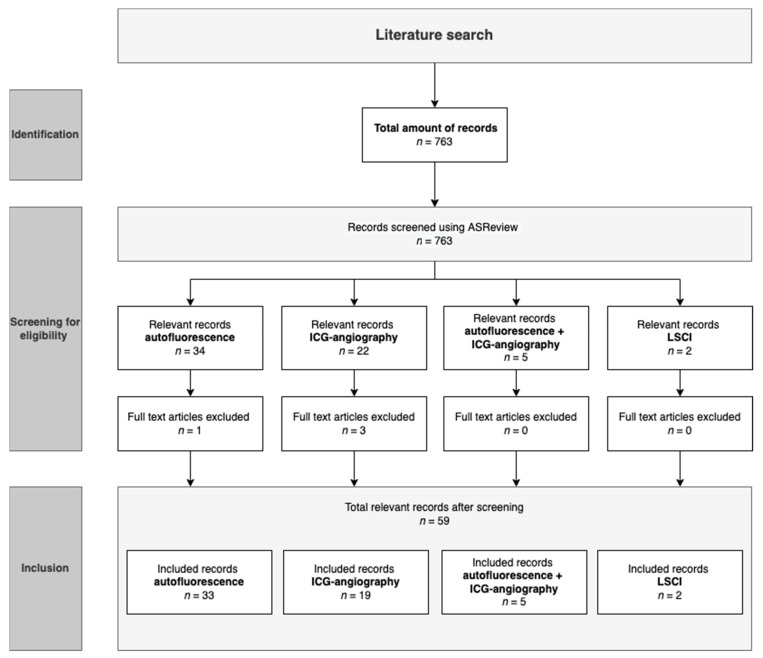
PUBMED, EMBASE and Web of Science were searched for studies written in the English language utilizing parathyroid autofluorescence, ICG-angiography, or LSCI during total thyroidectomy to support parathyroid gland identification or preservation. Case reports, reviews, meta-analyses, animal studies, and post-mortem studies were excluded after the title and abstract screening. The data of the studies were analyzed qualitatively, with a focus on the methodologies employed.
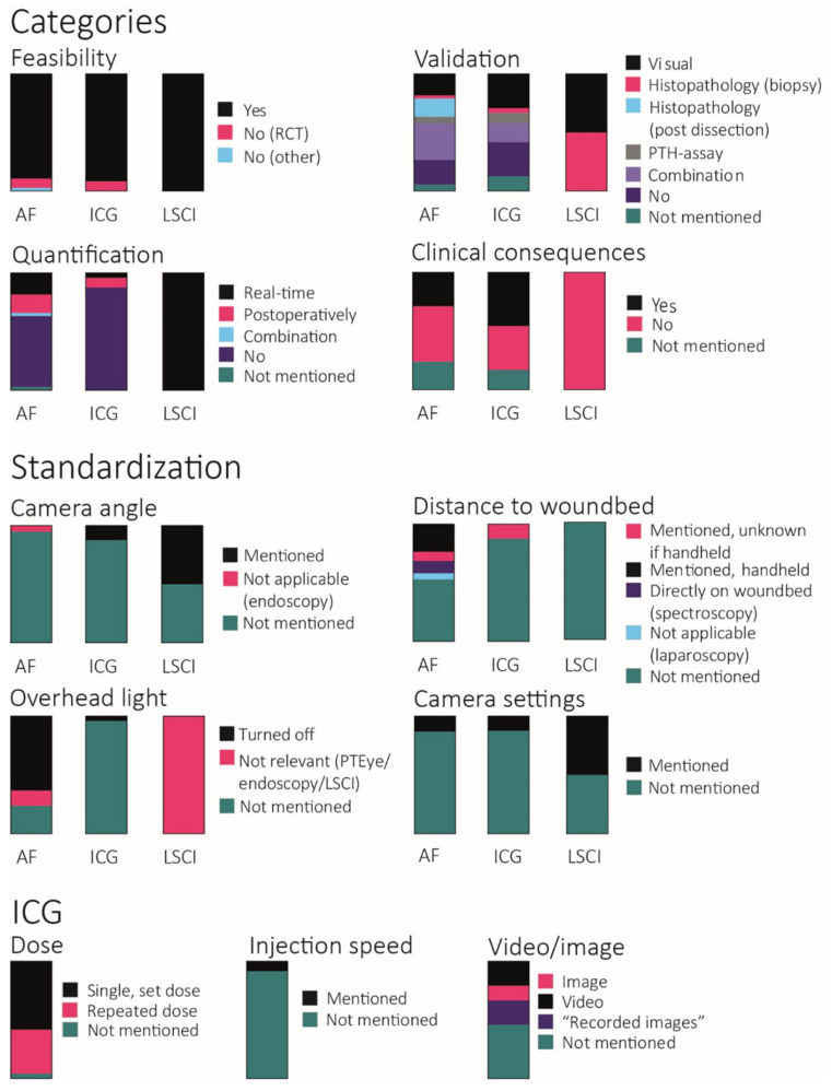
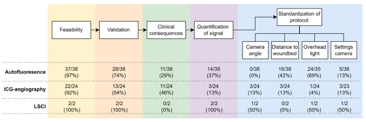
In total, 59 articles were included with a total of 6190 patients. Overall, 38 studies reported using parathyroid autofluorescence, 24 using ICG-angiography, and 2 using LSCI. The heterogeneity between the utilized methodology in the studies was large, and in particular, regarding study protocols, imaging techniques, and the standardization of the imaging protocol.
The diverse application of optical imaging techniques and a lack of standardization and quantification leads to heterogeneous conclusions regarding their clinical value. Worldwide consensus on imaging protocols is needed to establish the clinical utility of these techniques for parathyroid gland identification and preservation.
术后甲状旁腺功能减退是全甲状腺切除术后最常见的并发症。在过去几年中,光学成像技术,如甲状旁腺自发荧光、吲哚菁绿(ICG)血管造影和激光散斑对比成像(LSCI)已被用于在甲状腺手术中保护甲状旁腺。本研究概述了在全甲状腺切除术中用于识别和保护甲状旁腺的光学成像技术的应用方法。
检索PUBMED、EMBASE和科学网,查找在全甲状腺切除术中使用甲状旁腺自发荧光、ICG血管造影或LSCI以支持甲状旁腺识别或保护的英文研究。在标题和摘要筛选后,排除病例报告、综述、荟萃分析、动物研究和尸检研究。对研究数据进行定性分析,重点关注所采用的方法。
共纳入59篇文章,涉及6190例患者。总体而言,38项研究报告使用甲状旁腺自发荧光,24项使用ICG血管造影,2项使用LSCI。研究中所采用方法之间的异质性很大,特别是在研究方案、成像技术和成像方案的标准化方面。
光学成像技术的多样应用以及缺乏标准化和量化导致关于其临床价值的结论存在异质性。需要在全球范围内就成像方案达成共识,以确定这些技术在甲状旁腺识别和保护方面的临床实用性。