Department of Swine Diseases, National Veterinary Research Institute, Al. Partyzantów 57, 24-100 Puławy, Poland.
Department of Industrial and Environmental Microbiology, Institute of Biological Sciences, Maria Curie-Skłodowska University, 20-033 Lublin, Poland.
Molecules. 2022 Mar 21;27(6):2006. doi: 10.3390/molecules27062006.
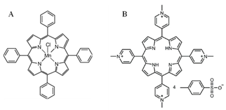
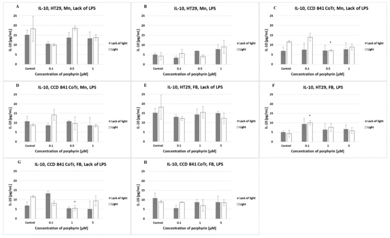
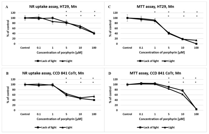
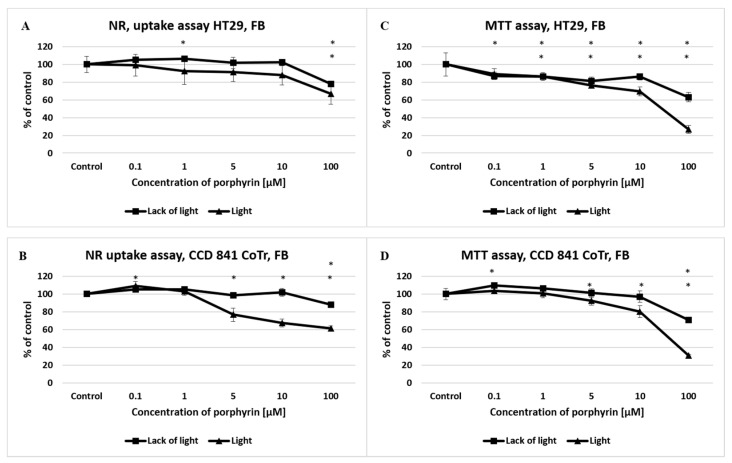
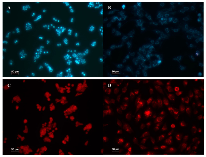
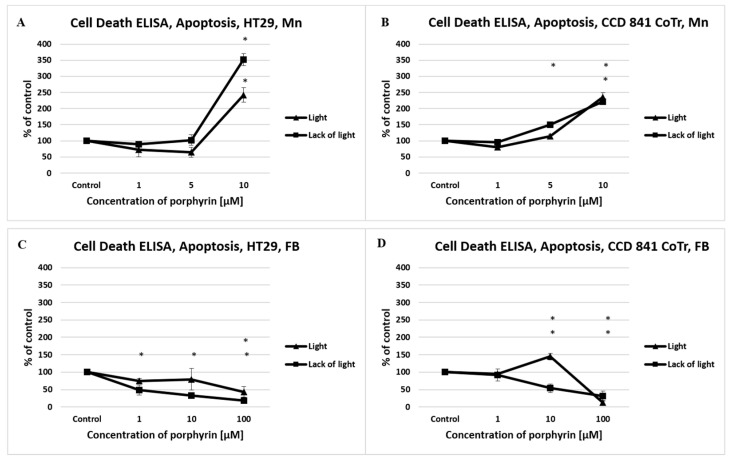
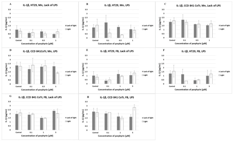
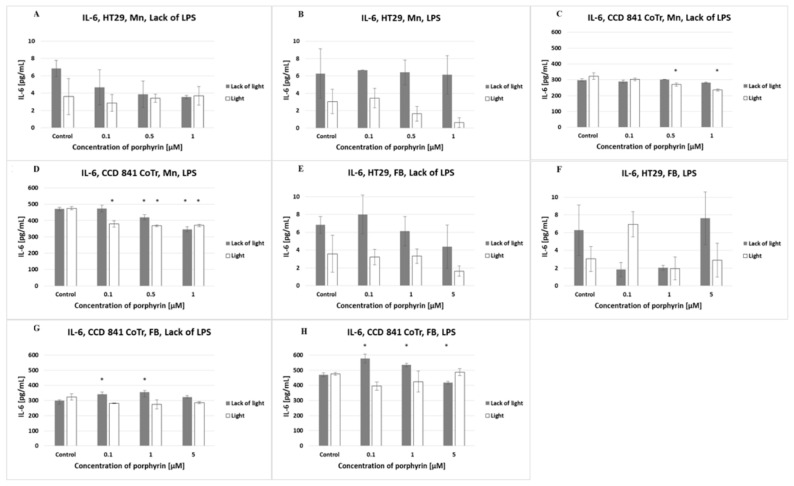
Standard in vitro analyses determining the activity of different compounds included in the chemotherapy of colon cancer are currently insufficient. New ideas, such as photodynamic therapy (PDT), may bring tangible benefits. The aim of this study was to show that the biological activity of selected free-base and manganese (III) metallated porphyrins differs in the limitation of colon cancer cell growth in vitro. White light irradiation was also hypothesized to initiate a photodynamic effect on tested porphyrins. Manganese porphyrin (>1 μM) significantly decreased the viability of the colon tumor and normal colon epithelial cells, both in light/lack of light conditions, while decreasing a free-base porphyrin after only 3 min of white light irradiation. Both porphyrins interacted with cytostatics in an antagonistic manner. The manganese porphyrin mainly induced apoptosis and necrosis in the tumor, and apoptosis in the normal cells, regardless of light exposure conditions. The free-base porphyrin conducted mainly apoptosis and autophagy. Normal and tumor cells released low levels of IL-1β and IL-10. Tumor cells released a low level of IL-6. Light conditions and porphyrins were influenced at the cytokine level. Tested manganese (III) metallated and free-base porphyrins differ in their activity against human colon cancer cells. The first showed no photodynamic, but a toxic activity, whereas the second expressed high photodynamic action. White light use may induce a photodynamic effect associated with porphyrins.
目前,用于确定结肠癌化疗中不同化合物活性的标准体外分析还不够充分。新的想法,如光动力疗法(PDT),可能会带来切实的好处。本研究旨在表明,选定的游离碱基和锰(III)金属化卟啉在体外限制结肠癌细胞生长方面的生物活性不同。还假设白光照射会引发测试卟啉的光动力效应。锰卟啉(>1 μM)在有光/无光条件下均显著降低结肠肿瘤和正常结肠上皮细胞的活力,而游离碱基卟啉仅在白光照射 3 分钟后才降低。两种卟啉均以拮抗方式与细胞抑制剂相互作用。锰卟啉主要在暴露于光和不暴露于光的条件下诱导肿瘤中的细胞凋亡和坏死,以及正常细胞中的细胞凋亡。游离碱基卟啉主要进行细胞凋亡和自噬。正常和肿瘤细胞释放低水平的 IL-1β 和 IL-10。肿瘤细胞释放低水平的 IL-6。光条件和卟啉在细胞因子水平上受到影响。测试的锰(III)金属化和游离碱基卟啉在其对人结肠癌细胞的活性方面存在差异。第一种卟啉没有表现出光动力活性,但具有毒性作用,而第二种卟啉则表现出高的光动力作用。白光的使用可能会引发与卟啉相关的光动力效应。