Gümrükçü Gülistan, Doğan Meryem, Gürsan Nilüfer, Boylu Barış, Ekren Erhan, Aker Fügen Vardar
University of Health Sciences, Hamidiye Faculty of Medicine, Department of Pathology, Haydarpaşa Numune Training and Research Hospital, Istanbul, Turkey.
Department of Pathology, Osmaniye Kadirli Public Hospital, Istanbul, Turkey.
J Cytol. 2022 Jan-Mar;39(1):30-36. doi: 10.4103/joc.joc_129_21. Epub 2022 Feb 17.
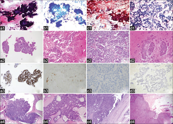
Diagnosis of papillary lesions of the breast by fine needle aspiration cytology (FNAC) is problematic. For this reason, it is situated in the indeterminate zone in classification systems.
To ascertain the accuracy of cytological diagnosis of papillary lesions in distinguishing papillary lesions from non-papillary lesions and to determine whether papillomas can be reliably distinguished from malignant papillary lesions by FNAC.
A total of 346 cases with the diagnoses of breast papillary lesions were selected among 5112 breast FNAC procedures performed in our center. One hundred and thirty-nine cases with excised lesions were included in this study, and their corresponding histology was reviewed.
Papillary lesion diagnosis was confirmed by histopathology in 103 (74.1%) of 139 patients. Cytology and histopathology results were not found to be compatible in 35 (25.2%) cases. The diagnostic accuracy of distinguishing papillary breast lesions as malignant or benign was assessed statistically. According to the cytology-histology comparison, one case was evaluated as false negative (FN) and twelve cases as false positive (FP). Overall accuracy, sensitivity, specificity, positive predictive value (PPV), and negative predictive value (NPV) of FNAC in distinguishing papillary lesions as benign or malignant were calculated as 87%, 97%, 83%, 72%, and 98%, respectively.
The diagnostic accuracy of papillary breast lesions classified by FNAC might be improved by careful evaluation together with cytological, radiological, and clinical findings (triple test). Cell block may allow more accurate evaluation of the papillary lesion and can be applied to immunohistochemical examination. It may also facilitate the differentiation of benign/malignant papillary lesions.
通过细针穿刺细胞学检查(FNAC)诊断乳腺乳头状病变存在问题。因此,在分类系统中它处于不确定区域。
确定FNAC对乳头状病变进行细胞学诊断以区分乳头状病变与非乳头状病变的准确性,并确定是否能通过FNAC可靠地区分乳头状瘤与恶性乳头状病变。
在本中心进行的5112例乳腺FNAC检查中,共选取了346例诊断为乳腺乳头状病变的病例。本研究纳入了139例有切除病变的病例,并对其相应的组织学进行了复查。
139例患者中,103例(74.1%)经组织病理学证实为乳头状病变诊断。35例(25.2%)病例的细胞学和组织病理学结果不相符。对区分乳腺乳头状病变为恶性或良性的诊断准确性进行了统计学评估。根据细胞学与组织学的比较,1例被评估为假阴性(FN),12例为假阳性(FP)。FNAC区分乳头状病变为良性或恶性的总体准确性、敏感性、特异性、阳性预测值(PPV)和阴性预测值(NPV)分别计算为87%、97%、83%、72%和98%。
通过结合细胞学、放射学和临床检查结果(三联检查)进行仔细评估,可能会提高FNAC对乳腺乳头状病变的诊断准确性。细胞块可使对乳头状病变的评估更准确,并可应用于免疫组化检查。它还可能有助于区分良性/恶性乳头状病变。