From the NAFLD Research Center, Department of Medicine (J.J., E.M., R.B., R.R.L., R.L.), Liver Imaging Group, Department of Radiology (A.S.B., K.J.F., C.B.S.), Department of Radiology (M.P.A.), and Division of Epidemiology, Department of Family and Preventive Medicine (R.L.), University of California at San Diego, ACTRI Building, 1W202, 9452 Medical Center Dr, La Jolla, CA 92037; Bioacoustics Research Laboratory, Department of Electrical and Computer Engineering (A.H., W.D.O.), and Department of Food Science and Human Nutrition (J.W.E.), University of Illinois at Urbana-Champaign, Urbana, Ill.
Radiology. 2022 Jul;304(1):75-82. doi: 10.1148/radiol.211131. Epub 2022 Mar 29.
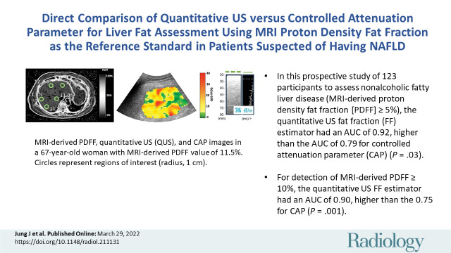
Background MRI-derived proton density fat fraction (PDFF) is an accurate, reliable, and safe biologic marker for use in the noninvasive diagnosis of hepatic steatosis in patients with nonalcoholic fatty liver disease (NAFLD). Because of the cost and limited availability of MRI, it is necessary to develop an accurate method to diagnose NAFLD with potential point-of-care access. Purpose To compare the diagnostic accuracy of the quantitative US (QUS) fat fraction (FF) estimator with that of the controlled attenuation parameter (CAP) in the diagnosis of NAFLD using contemporaneous MRI-derived PDFF as the reference standard. Materials and Methods Participants with or suspected of having NAFLD were prospectively recruited at the NAFLD Research Center between July 2015 and July 2019. All participants underwent MRI-derived PDFF measurement, transient elastography with CAP measurement, and QUS. QUS FF was derived using computed QUS parameters from the acquired radiofrequency US data using a calibrated reference phantom. The area under the receiver operating characteristic curve (AUC) was calculated to assess the accuracy of QUS FF and CAP in the diagnosis of hepatic steatosis (defined as MRI-derived PDFF ≥ 5%). AUCs were compared using the DeLong test. Results A total of 123 participants were included (mean age, 52 years ± 13 [SD]; 67 [54%] women). Of these participants, 100 (81%) had MRI-derived PDFF of 5% or more. QUS FF had a significantly higher AUC for diagnosis of NAFLD than did CAP (0.92 [95% CI: 0.87, 0.98] vs 0.79 [95% CI: 0.67, 0.90], = .03). QUS FF had a sensitivity of 98% (98 of 100) and a specificity of 48% (11 of 23). CAP had a sensitivity of 87% (87 of 100) and a specificity of 57% (13 of 23). Conclusion The quantitative US fat fraction estimator is more accurate than the controlled attenuation parameter in the diagnosis of hepatic steatosis in patients with or suspected of having nonalcoholic fatty liver disease. © RSNA, 2022 See also the editorial by Ito in this issue.
背景 基于 MRI 的质子密度脂肪分数(PDFF)是一种准确、可靠且安全的生物学标志物,可用于非酒精性脂肪性肝病(NAFLD)患者的非侵入性肝脂肪变性诊断。由于 MRI 的成本和可用性有限,因此需要开发一种准确的方法,以便在具备潜在即时检测条件的情况下诊断 NAFLD。目的 本研究旨在比较定量超声(QUS)脂肪分数(FF)估测值与受控衰减参数(CAP)在以同期 MRI 衍生 PDFF 为参考标准的 NAFLD 诊断中的诊断准确性。材料与方法 2015 年 7 月至 2019 年 7 月,前瞻性地在 NAFLD 研究中心招募了疑似或确诊为 NAFLD 的患者。所有参与者均接受了 MRI 衍生 PDFF 测量、CAP 测量和 QUS 检查。QUS FF 是通过使用校准参考体模从采集的射频 US 数据中计算出的计算 QUS 参数得出的。采用受试者工作特征曲线(ROC)下面积(AUC)评估 QUS FF 和 CAP 对肝脂肪变性(定义为 MRI 衍生 PDFF≥5%)的诊断准确性。采用 DeLong 检验比较 AUC。结果 共纳入 123 名参与者(平均年龄,52 岁±13[标准差];67[54%]为女性)。其中,100 名(81%)参与者的 MRI 衍生 PDFF 为 5%或更高。与 CAP 相比,QUS FF 对诊断 NAFLD 的 AUC 显著更高(0.92[95%CI:0.87,0.98] vs 0.79[95%CI:0.67,0.90], =.03)。QUS FF 的敏感度为 98%(100 例中的 98 例),特异度为 48%(23 例中的 11 例)。CAP 的敏感度为 87%(100 例中的 87 例),特异度为 57%(23 例中的 13 例)。结论 在诊断疑似或确诊为 NAFLD 的患者的肝脂肪变性方面,定量 US 脂肪分数估测值比 CAP 更准确。©2022 RSNA。另见本期 Ito 编辑述评。