Ros Tomas, Michela Abele, Mayer Anaïs, Bellmann Anne, Vuadens Philippe, Zermatten Victorine, Saj Arnaud, Vuilleumier Patrik
Department of Neuroscience, University of Geneva, Geneva, Switzerland.
CIBM Center for Biomedical Imaging, Geneva University Hospitals, Geneva, Switzerland.
Netw Neurosci. 2022 Feb 1;6(1):69-89. doi: 10.1162/netn_a_00210. eCollection 2022 Feb.
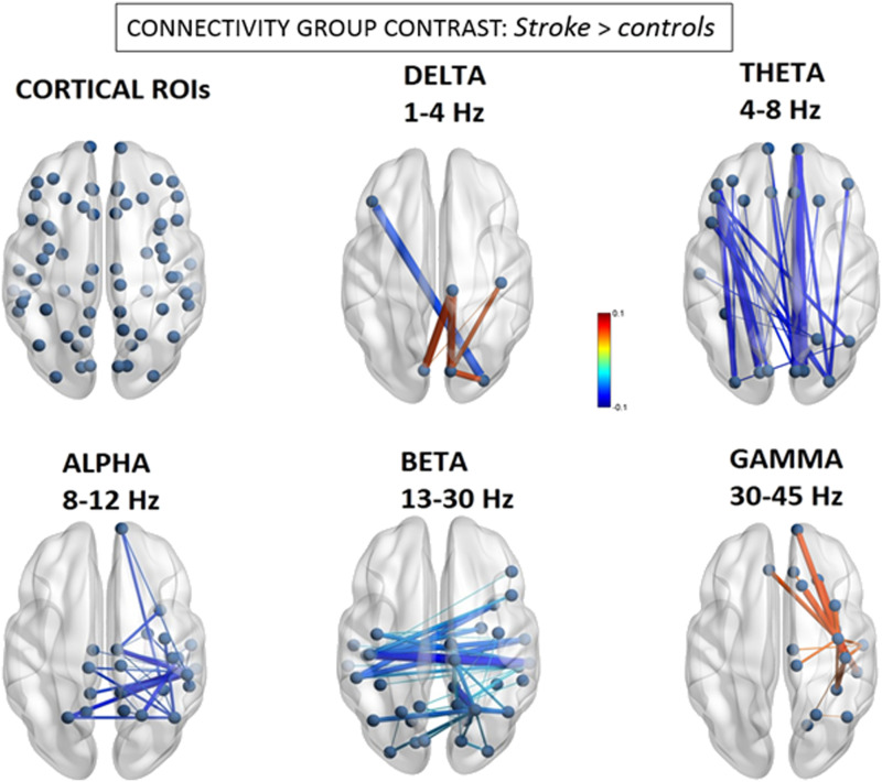
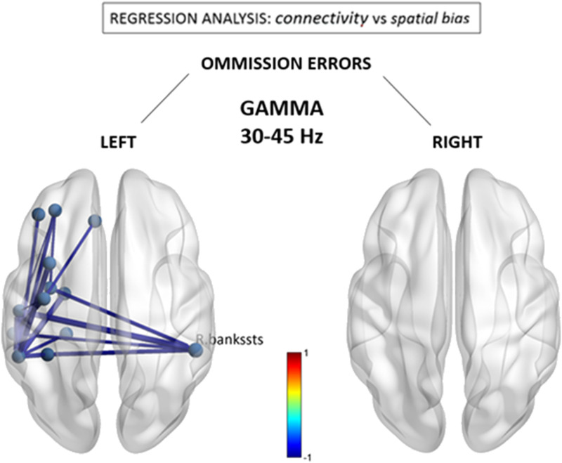
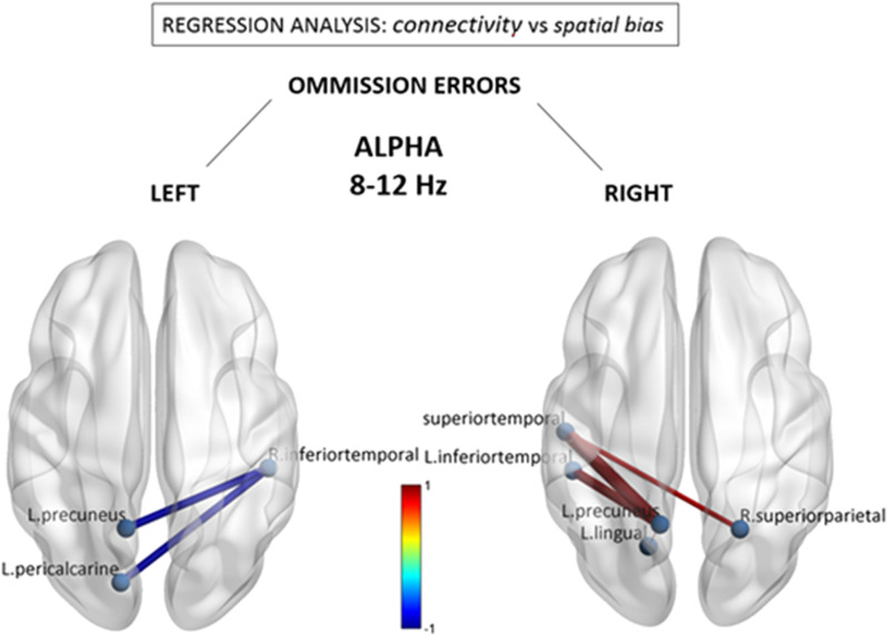
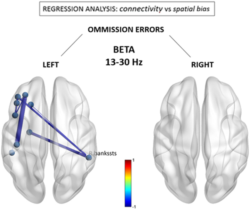
Stroke frequently produces attentional dysfunctions including symptoms of hemispatial neglect, which is characterized by a breakdown of awareness for the contralesional hemispace. Recent studies with functional MRI (fMRI) suggest that hemineglect patients display abnormal - and functional connectivity. However, since stroke is a vascular disorder and fMRI signals remain sensitive to nonneuronal (i.e., vascular) coupling, more direct demonstrations of neural network dysfunction in hemispatial neglect are warranted. Here, we utilize electroencephalogram (EEG) source imaging to uncover differences in resting-state network organization between patients with right hemispheric stroke ( = 15) and age-matched, healthy controls ( = 27), and determine the relationship between hemineglect symptoms and brain network organization. We estimated - and differences in cortical communication by calculating the spectral power and amplitude envelope correlations of narrow-band EEG oscillations. We first observed focal frequency-slowing within the right posterior cortical regions, reflected in relative delta/theta power increases and alpha/beta/gamma decreases. Secondly, nodes within the right temporal and parietal cortex consistently displayed anomalous intra- and interhemispheric coupling, stronger in delta and gamma bands, and weaker in theta, alpha, and beta bands. Finally, a significant association was observed between the severity of left-hemispace search deficits (e.g., cancellation test omissions) and reduced functional connectivity within the alpha and beta bands. In sum, our novel results validate the hypothesis of large-scale cortical network disruption following stroke and reinforce the proposal that abnormal brain oscillations may be intimately involved in the pathophysiology of visuospatial neglect.
中风经常会导致注意力功能障碍,包括半侧空间忽视症状,其特征是对患侧半空间的意识丧失。最近的功能磁共振成像(fMRI)研究表明,半侧忽视患者表现出异常的功能连接。然而,由于中风是一种血管疾病,且fMRI信号对非神经元(即血管)耦合仍很敏感,因此有必要更直接地证明半侧空间忽视中神经网络功能障碍。在这里,我们利用脑电图(EEG)源成像来揭示右半球中风患者(n = 15)与年龄匹配的健康对照组(n = 27)在静息状态网络组织上的差异,并确定半侧忽视症状与脑网络组织之间的关系。我们通过计算窄带EEG振荡的频谱功率和幅度包络相关性来估计皮质通信的差异。我们首先观察到右后皮质区域出现局灶性频率减慢,表现为相对δ/θ功率增加,α/β/γ功率降低。其次,右侧颞叶和顶叶皮质内的节点始终表现出异常的半球内和半球间耦合,在δ和γ波段更强,在θ、α和β波段较弱。最后,观察到左半空间搜索缺陷的严重程度(如取消测试遗漏)与α和β波段内功能连接减少之间存在显著关联。总之,我们的新结果验证了中风后大规模皮质网络破坏的假设,并强化了异常脑振荡可能与视觉空间忽视的病理生理学密切相关的观点。