Rusiniak Mateusz, Bornfleth Harald, Cho Jae-Hyun, Wolak Tomasz, Ille Nicole, Berg Patrick, Scherg Michael
Research Department, BESA GmbH, Gräfelfing, Germany.
Bioimaging Research Center, World Hearing Center of the Institute of Physiology and Pathology of Hearing, Warsaw, Poland.
Front Neurosci. 2022 Mar 10;16:842420. doi: 10.3389/fnins.2022.842420. eCollection 2022.
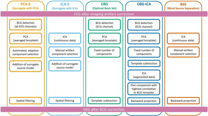
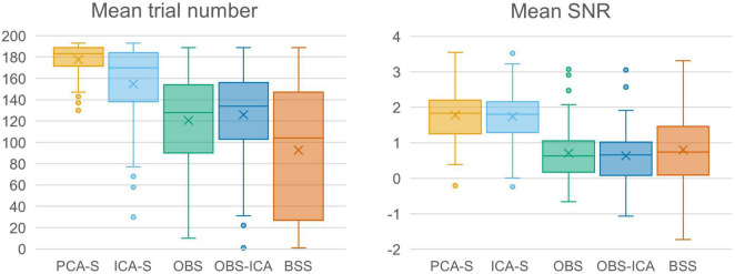
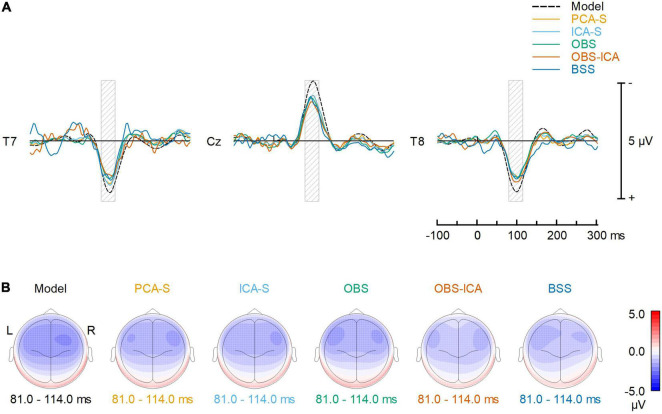
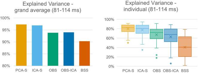
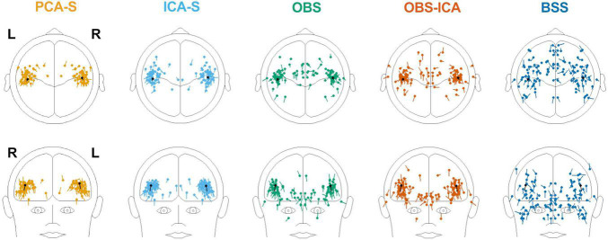
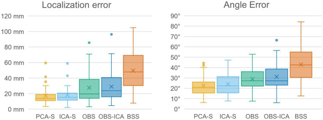
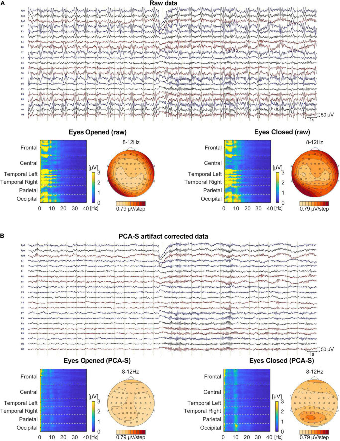
For the analysis of simultaneous EEG-fMRI recordings, it is vital to use effective artifact removal tools. This applies in particular to the ballistocardiogram (BCG) artifact which is difficult to remove without distorting signals of interest related to brain activity. Here, we documented the use of surrogate source models to separate the artifact-related signals from brain signals with minimal distortion of the brain activity of interest. The artifact topographies used for surrogate separation were created automatically using principal components analysis (PCA-S) or by manual selection of artifact components utilizing independent components analysis (ICA-S). Using real resting-state data from 55 subjects superimposed with simulated auditory evoked potentials (AEP), both approaches were compared with three established BCG artifact removal methods: Blind Source Separation (BSS), Optimal Basis Set (OBS), and a mixture of both (OBS-ICA). Each method was evaluated for its applicability for ERP and source analysis using the following criteria: the number of events surviving artifact threshold scans, signal-to-noise ratio (SNR), error of source localization, and signal variance explained by the dipolar model. Using these criteria, PCA-S and ICA-S fared best overall, with highly significant differences to the established methods, especially in source localization. The PCA-S approach was also applied to a single subject Berger experiment performed in the MRI scanner. Overall, the removal of BCG artifacts by the surrogate methods provides a substantial improvement for the analysis of simultaneous EEG-fMRI data compared to the established methods.
对于同步脑电图-功能磁共振成像(EEG-fMRI)记录的分析,使用有效的伪迹去除工具至关重要。这尤其适用于心冲击图(BCG)伪迹,在不扭曲与大脑活动相关的感兴趣信号的情况下,很难将其去除。在此,我们记录了使用替代源模型将与伪迹相关的信号与大脑信号分离,同时使感兴趣的大脑活动的失真最小化。用于替代分离的伪迹地形图是使用主成分分析自动创建的(PCA-S),或者通过利用独立成分分析手动选择伪迹成分来创建(ICA-S)。使用来自55名受试者的真实静息状态数据,并叠加模拟听觉诱发电位(AEP),将这两种方法与三种已确立的BCG伪迹去除方法进行比较:盲源分离(BSS)、最优基集(OBS)以及两者的混合(OBS-ICA)。使用以下标准评估每种方法对事件相关电位(ERP)和源分析的适用性:在伪迹阈值扫描后幸存的事件数量、信噪比(SNR)、源定位误差以及偶极模型解释的信号方差。使用这些标准,PCA-S和ICA-S总体表现最佳,与已确立的方法有显著差异,尤其是在源定位方面。PCA-S方法还应用于在MRI扫描仪中进行的单个受试者的贝格尔实验。总体而言,与已确立的方法相比,通过替代方法去除BCG伪迹为同步EEG-fMRI数据的分析提供了实质性的改进。