Egorova-Brumley Natalia, Khlif Mohamed Salah, Werden Emilio, Bird Laura J, Brodtmann Amy
The Florey Institute of Neuroscience and Mental Health, Melbourne, Australia.
The University of Melbourne, Melbourne, Australia.
Brain Commun. 2022 Mar 17;4(2):fcac061. doi: 10.1093/braincomms/fcac061. eCollection 2022.
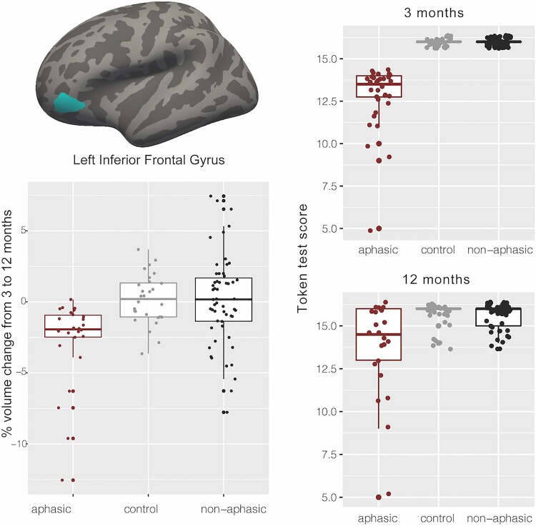
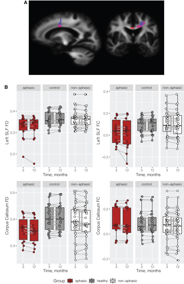
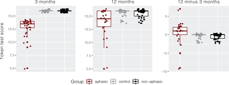
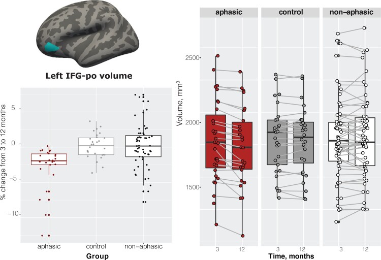
Dynamic whole-brain changes occur following stroke, and not just in association with recovery. We tested the hypothesis that the presence of a specific behavioural deficit after stroke would be associated with structural decline (atrophy) in the brain regions supporting the affected function, by examining language deficits post-stroke. We quantified whole-brain structural volume changes longitudinally (3-12 months) in stroke participants with ( = 32) and without aphasia ( = 59) as assessed by the Token Test at 3 months post-stroke, compared with a healthy control group ( = 29). While no significant difference in language decline rates (change in Token Test scores from 3 to 12 months) was observed between groups and some participants in the aphasic group improved their scores, stroke participants with aphasia symptoms at 3 months showed significant atrophy (>2%, = 0.0001) of the left inferior frontal gyrus not observed in either healthy control or non-aphasic groups over the 3-12 months period. We found significant group differences in the inferior frontal gyrus volume, accounting for age, sex, stroke severity at baseline, education and total intracranial volume (Bonferroni-corrected = 0.0003). In a subset of participants (aphasic = 14, non-aphasic = 36, and healthy control = 25) with available diffusion-weighted imaging data, we found significant atrophy in the corpus callosum and the left superior longitudinal fasciculus in the aphasic compared with the healthy control group. Language deficits at 3 months post-stroke are associated with accelerated structural decline specific to the left inferior frontal gyrus, highlighting that known functional brain reorganization underlying behavioural improvement may occur in parallel with atrophy of brain regions supporting the language function.
中风后会出现全脑动态变化,且不仅仅与恢复过程相关。我们通过研究中风后的语言缺陷,验证了这样一个假设:中风后特定行为缺陷的存在会与支持受影响功能的脑区结构衰退(萎缩)相关。我们纵向(3 - 12个月)量化了中风参与者(根据中风后3个月的代币测试评估,有失语症的n = 32,无失语症的n = 59)与健康对照组(n = 29)的全脑结构体积变化。虽然各小组之间在语言衰退率(代币测试分数从3个月到12个月的变化)上未观察到显著差异,且失语症组的一些参与者分数有所提高,但在3个月时有失语症症状的中风参与者在3 - 12个月期间左额下回出现了显著萎缩(>2%,p = 0.0001),这在健康对照组或无失语症组中均未观察到。我们发现额下回体积存在显著的组间差异,该差异在考虑了年龄、性别、基线中风严重程度、教育程度和总颅内体积后仍然显著(Bonferroni校正p = 0.0003)。在一部分有可用扩散加权成像数据的参与者中(失语症患者n = 14,非失语症患者n = 36,健康对照组n = 25),我们发现与健康对照组相比,失语症患者的胼胝体和左侧上纵束出现了显著萎缩。中风后3个月的语言缺陷与左额下回特有的结构加速衰退相关,这突出表明行为改善背后已知的脑功能重组可能与支持语言功能的脑区萎缩同时发生。