Human Biology Division, Fred Hutchinson Cancer Research Center, Seattle, Washington, United States of America.
Medical Scientist Training Program, University of Washington, Seattle, Washington, United States of America.
PLoS Pathog. 2022 Apr 11;18(4):e1010155. doi: 10.1371/journal.ppat.1010155. eCollection 2022 Apr.
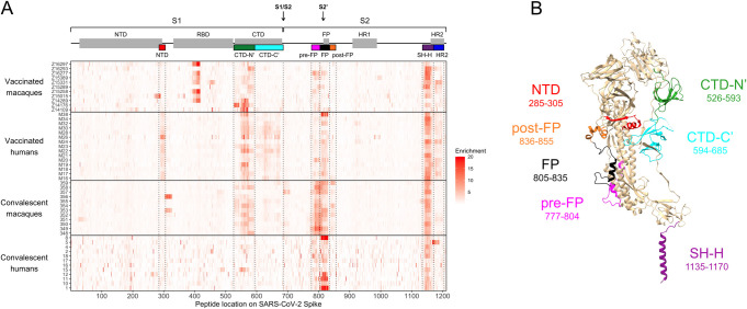
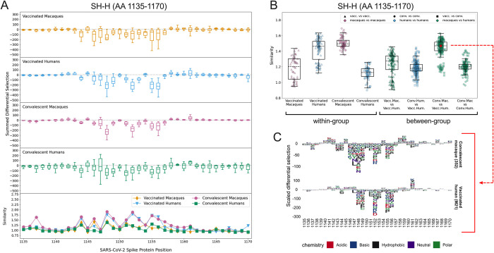
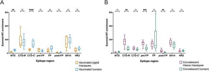
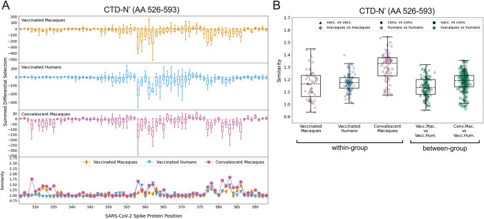
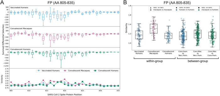
Macaques are a commonly used model for studying immunity to human viruses, including for studies of SARS-CoV-2 infection and vaccination. However, it is unknown whether macaque antibody responses resemble the response in humans. To answer this question, we employed a phage-based deep mutational scanning approach (Phage-DMS) to compare which linear epitopes are targeted on the SARS-CoV-2 Spike protein in convalescent humans, convalescent (re-infected) rhesus macaques, mRNA-vaccinated humans, and repRNA-vaccinated pigtail macaques. We also used Phage-DMS to determine antibody escape pathways within each epitope, enabling a granular comparison of antibody binding specificities at the locus level. Overall, we identified some common epitope targets in both macaques and humans, including in the fusion peptide (FP) and stem helix-heptad repeat 2 (SH-H) regions. Differences between groups included a response to epitopes in the N-terminal domain (NTD) and C-terminal domain (CTD) in vaccinated humans but not vaccinated macaques, as well as recognition of a CTD epitope and epitopes flanking the FP in convalescent macaques but not convalescent humans. There was also considerable variability in the escape pathways among individuals within each group. Sera from convalescent macaques showed the least variability in escape overall and converged on a common response with vaccinated humans in the SH-H epitope region, suggesting highly similar antibodies were elicited. Collectively, these findings suggest that the antibody response to SARS-CoV-2 in macaques shares many features with humans, but with substantial differences in the recognition of certain epitopes and considerable individual variability in antibody escape profiles, suggesting a diverse repertoire of antibodies that can respond to major epitopes in both humans and macaques. Differences in macaque species and exposure type may also contribute to these findings.
猕猴常用于研究人类病毒的免疫反应,包括 SARS-CoV-2 感染和疫苗接种的研究。然而,猕猴的抗体反应是否与人类相似尚不清楚。为了回答这个问题,我们采用噬菌体展示深度突变扫描(Phage-DMS)方法,比较恢复期人类、恢复期(再次感染)恒河猴、mRNA 疫苗接种人类和 repRNA 疫苗接种猪尾猕猴体内针对 SARS-CoV-2 刺突蛋白的线性表位。我们还使用 Phage-DMS 来确定每个表位内的抗体逃逸途径,从而能够在基因座水平上对抗体结合特异性进行精细比较。总体而言,我们在猕猴和人类中都鉴定到了一些共同的表位靶标,包括融合肽(FP)和茎螺旋-七肽重复 2(SH-H)区。各组之间的差异包括接种疫苗的人类对 N 端结构域(NTD)和 C 端结构域(CTD)中的表位的反应,但接种疫苗的猕猴没有;以及在恢复期猕猴中识别 CTD 表位和 FP 侧翼表位,但在恢复期人类中没有。每个组内个体的逃逸途径也存在相当大的差异。恢复期猕猴的血清总体上表现出最小的逃逸变异性,在 SH-H 表位区域与接种疫苗的人类趋同,表明诱导出高度相似的抗体。总的来说,这些发现表明,猕猴对 SARS-CoV-2 的抗体反应与人类有许多共同特征,但在某些表位的识别和抗体逃逸谱的个体变异性方面存在很大差异,这表明在人类和猕猴中都能对主要表位产生反应的抗体具有多样性。猕猴物种和暴露类型的差异也可能导致这些发现。